 Movie
Movie Controller

[English] 日本語
 Yorodumi
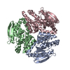
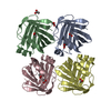
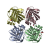
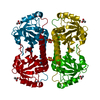
- PDB-4hc8: CRYSTAL STRUCTURE OF PROBABLE ENOYL-CoA HYDRATASE ECHA3 (Rv0632c,... -
+ Open data

- Basic information
| Entry | Database: PDB / ID: 4hc8 | ||||||
|---|---|---|---|---|---|---|---|
| Title | CRYSTAL STRUCTURE OF PROBABLE ENOYL-CoA HYDRATASE ECHA3 (Rv0632c, NYSGRC-019494) from Mycobacterium Tuberculosis H37Rv | ||||||
 Components Components | Enoyl-CoA hydratase/isomerase family protein | ||||||
 Keywords Keywords | LYASE / ECHA3 / UNSATURATED ACYL-CoA HYDRATASE / CROTONASE / Structural genomics / NYSGRC / PSI / New York Structural Genomics Research Consortium / PSI-Biology | ||||||
| Function / homology |  Function and homology information Function and homology informationenoyl-CoA hydratase / enoyl-CoA hydratase activity / fatty acid beta-oxidation / plasma membrane / cytosol Similarity search - Function | ||||||
| Biological species |   Mycobacterium tuberculosis (bacteria) Mycobacterium tuberculosis (bacteria) | ||||||
| Method |  X-RAY DIFFRACTION / SYNCHROTRON / SAD / Resolution: 2.51 Å X-RAY DIFFRACTION / SYNCHROTRON / SAD / Resolution: 2.51 Å | ||||||
 Authors Authors | Sampathkumar, P. / Almo, S.C. / New York Structural Genomics Research Consortium (NYSGRC) | ||||||
 Citation Citation | Journal: To be Published Title: Crystal structure of probable enoyl-CoA hydratase ECHA3 (Rv0632c, NYSGRC-019494) from Mycobacterium Tuberculosis H37Rv Authors: Sampathkumar, P. / Ahmed, M. / Banu, N. / Bhosle, R. / Bonanno, J. / Chamala, S. / Chowdhury, S. / Fiser, A. / Gizzi, A. / Glenn, A.S. / Hammonds, J. / Hillerich, B. / Khafizov, K. / ...Authors: Sampathkumar, P. / Ahmed, M. / Banu, N. / Bhosle, R. / Bonanno, J. / Chamala, S. / Chowdhury, S. / Fiser, A. / Gizzi, A. / Glenn, A.S. / Hammonds, J. / Hillerich, B. / Khafizov, K. / Lafleur, J. / Love, J.D. / Stead, M. / Seidel, R. / Toro, R. / Almo, S.C. | ||||||
| History |
|
- Structure visualization
| Structure viewer | Molecule: MolmilJmol/JSmol |
|---|
- Downloads & links
-Download
| PDBx/mmCIF format | 4hc8.cif.gz | 103.1 KB | Display | PDBx/mmCIF format |
|---|---|---|---|---|
| PDB format | pdb4hc8.ent.gz | 80.9 KB | Display | PDB format |
| PDBx/mmJSON format | 4hc8.json.gz | Tree view | PDBx/mmJSON format | |
| Others |  Other downloads Other downloads |
-Validation report
| Arichive directory | https://data.pdbj.org/pub/pdb/validation_reports/hc/4hc8ftp://data.pdbj.org/pub/pdb/validation_reports/hc/4hc8 | HTTPS FTP |
|---|
-Related structure data
| Similar structure data | |
|---|---|
| Other databases |


-Links
 PDBj
PDBj- Assembly
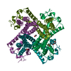
| Deposited unit | 
| ||||||||||||||||||||||||
|---|---|---|---|---|---|---|---|---|---|---|---|---|---|---|---|---|---|---|---|---|---|---|---|---|---|
| 1 | 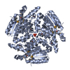
| ||||||||||||||||||||||||
| 2 | 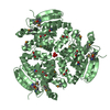
| ||||||||||||||||||||||||
| Unit cell |
| ||||||||||||||||||||||||
| Components on special symmetry positions |
|
-Components
| #1: Protein | Mass: 27856.297 Da / Num. of mol.: 2 Source method: isolated from a genetically manipulated source Source: (gene. exp.) Mycobacterium tuberculosis (bacteria) / Strain: H37Rv / Gene: echA3, MT0660, Rv0632c / Plasmid: CHS30 / Production host: #2: Chemical | ChemComp-SO4 /   Mass: 96.063 Da / Num. of mol.: 4 / Source method: obtained synthetically / Formula: SO4 Mass: 96.063 Da / Num. of mol.: 4 / Source method: obtained synthetically / Formula: SO4#3: Chemical |   Mass: 62.068 Da / Num. of mol.: 2 / Source method: obtained synthetically / Formula: C2H6O2 Mass: 62.068 Da / Num. of mol.: 2 / Source method: obtained synthetically / Formula: C2H6O2#4: Water | ChemComp-HOH / |  Mass: 18.015 Da / Num. of mol.: 67 / Source method: isolated from a natural source / Formula: H2O Mass: 18.015 Da / Num. of mol.: 67 / Source method: isolated from a natural source / Formula: H2OHas protein modification | Y | |
|---|
-Experimental details
-Experiment
| Experiment | Method: X-RAY DIFFRACTION / Number of used crystals: 1 |
|---|
- Sample preparation
| Crystal | Density Matthews: 2.78 Å3/Da / Density % sol: 55.73 % |
|---|---|
| Crystal grow | Temperature: 298 K / Method: vapor diffusion, sitting drop / pH: 6.5 Details: Protein (20 mM Hepes, pH 7.5, 150 mM NaCl, 10% glycerol, Reservoir (Crystal Screen F11: 1.6 M Ammonium sulfate, 0.1 M MES monohydrate pH 6.5, 10% v/v 1,4-Dioxane), Cryoprotection (30% ...Details: Protein (20 mM Hepes, pH 7.5, 150 mM NaCl, 10% glycerol, Reservoir (Crystal Screen F11: 1.6 M Ammonium sulfate, 0.1 M MES monohydrate pH 6.5, 10% v/v 1,4-Dioxane), Cryoprotection (30% Ethylene glycol), VAPOR DIFFUSION, SITTING DROP, temperature 298K |
-Data collection
| Diffraction | Mean temperature: 100 K |
|---|---|
| Diffraction source | Source: SYNCHROTRON / Site: NSLS  / Beamline: X29A / Wavelength: 0.9792 Å / Beamline: X29A / Wavelength: 0.9792 Å |
| Detector | Type: ADSC QUANTUM 315 / Detector: CCD / Date: Feb 1, 2012 / Details: MIRRORS |
| Radiation | Monochromator: Si(111) / Protocol: SINGLE WAVELENGTH / Monochromatic (M) / Laue (L): M / Scattering type: x-ray |
| Radiation wavelength | Wavelength: 0.9792 Å / Relative weight: 1 |
| Reflection | Resolution: 2.51→50 Å / Num. all: 21213 / Num. obs: 21213 / % possible obs: 100 % / Observed criterion σ(F): 0 / Observed criterion σ(I): 0 / Redundancy: 21.9 % / Biso Wilson estimate: 62 Å2 / Rsym value: 0.1 / Net I/σ(I): 27.07 |
| Reflection shell | Resolution: 2.51→2.55 Å / Redundancy: 21 % / Mean I/σ(I) obs: 4.7 / Num. unique all: 1054 / Rsym value: 0.977 / % possible all: 100 |
- Processing
| Software |
| ||||||||||||||||||||||||||||||||||||||||||||||||||||||||||||
|---|---|---|---|---|---|---|---|---|---|---|---|---|---|---|---|---|---|---|---|---|---|---|---|---|---|---|---|---|---|---|---|---|---|---|---|---|---|---|---|---|---|---|---|---|---|---|---|---|---|---|---|---|---|---|---|---|---|---|---|---|---|
| Refinement | Method to determine structure: SAD Starting model: Built using Buccaneer Resolution: 2.51→41.43 Å / Cor.coef. Fo:Fc: 0.96 / Cor.coef. Fo:Fc free: 0.934 / Occupancy max: 1 / Occupancy min: 0.33 / SU B: 8.841 / SU ML: 0.195 / Cross valid method: THROUGHOUT / σ(F): 0 / ESU R: 0.372 / ESU R Free: 0.27 / Stereochemistry target values: MAXIMUM LIKELIHOOD Details: HYDROGENS HAVE BEEN ADDED IN THE RIDING POSITIONS U VALUES : REFINED INDIVIDUALLY
| ||||||||||||||||||||||||||||||||||||||||||||||||||||||||||||
| Solvent computation | Ion probe radii: 0.8 Å / Shrinkage radii: 0.8 Å / VDW probe radii: 1.2 Å / Solvent model: MASK | ||||||||||||||||||||||||||||||||||||||||||||||||||||||||||||
| Displacement parameters | Biso max: 127.17 Å2 / Biso mean: 58.8883 Å2 / Biso min: 28.54 Å2
| ||||||||||||||||||||||||||||||||||||||||||||||||||||||||||||
| Refinement step | Cycle: LAST / Resolution: 2.51→41.43 Å
| ||||||||||||||||||||||||||||||||||||||||||||||||||||||||||||
| Refine LS restraints |
| ||||||||||||||||||||||||||||||||||||||||||||||||||||||||||||
| LS refinement shell | Resolution: 2.513→2.578 Å / Total num. of bins used: 20
|
