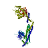
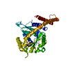
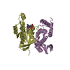
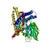
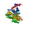
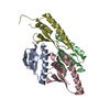
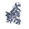
登録情報 データベース : PDB / ID : 4h1uタイトル Nucleotide-free human dynamin-1-like protein GTPase-GED fusion Dynamin-1-like protein キーワード / / 機能・相同性 分子機能 ドメイン・相同性 構成要素
/ / / / / / / / / / / / / / / / / / / / / / / / / / / / / / / / / / / / / / / / / / / / / / / / / / / / / / / / / / / / / / / / / / / / / / / / / / / / / / / / / / / / 生物種 Homo sapiens (ヒト)手法 / / / 解像度 : 2.3 Å データ登録者 Wenger, J. / Klinglmayr, E. / Puehringer, S. / Goettig, P. ジャーナル : Plos One / 年 : 2013タイトル : Functional Mapping of Human Dynamin-1-Like GTPase Domain Based on X-ray Structure Analyses.著者 : Wenger, J. / Klinglmayr, E. / Frohlich, C. / Eibl, C. / Gimeno, A. / Hessenberger, M. / Puehringer, S. / Daumke, O. / Goettig, P. 履歴 登録 2012年9月11日 登録サイト / 処理サイト 改定 1.0 2013年8月21日 Provider / タイプ 改定 1.1 2013年9月11日 Group 改定 1.2 2017年8月9日 Group / Source and taxonomy / カテゴリ / software改定 1.3 2023年9月13日 Group Data collection / Database references ... Data collection / Database references / Derived calculations / Refinement description カテゴリ chem_comp_atom / chem_comp_bond ... chem_comp_atom / chem_comp_bond / database_2 / pdbx_initial_refinement_model / struct_ref_seq_dif / struct_site Item _database_2.pdbx_DOI / _database_2.pdbx_database_accession ... _database_2.pdbx_DOI / _database_2.pdbx_database_accession / _struct_ref_seq_dif.details / _struct_site.pdbx_auth_asym_id / _struct_site.pdbx_auth_comp_id / _struct_site.pdbx_auth_seq_id
すべて表示 表示を減らす
 ムービー
ムービー コントローラー


 データを開く
 基本情報
基本情報 要素
要素 キーワード
キーワード 機能・相同性情報
 Homo sapiens (ヒト)
 X線回折 /
  データ登録者
 引用
引用 構造の表示
構造の表示 ダウンロードとリンク
 その他のダウンロード


 PDBj
PDBj


 集合体
集合体


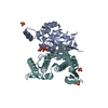
 分子量: 189.100 Da / 分子数: 1 / 由来タイプ: 合成 / 式: C6H5O7
 分子量: 18.015 Da / 分子数: 129 / 由来タイプ: 天然 / 式: H2O
 試料調製
試料調製 / ビームライン: 14.1 / 波長: 0.91841 Å
 解析
解析