 Movie
Movie Controller

[English] 日本語
 Yorodumi
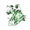
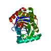
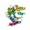
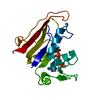
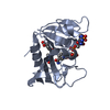
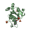
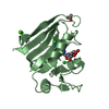
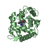
- PDB-4gvw: Three-dimensional structure of the de novo designed serine hydrol... -
+ Open data

- Basic information
| Entry | Database: PDB / ID: 4gvw | ||||||
|---|---|---|---|---|---|---|---|
| Title | Three-dimensional structure of the de novo designed serine hydrolase 2bfq_3, Northeast Structural Genomics Consortium (NESG) Target OR248 | ||||||
 Components Components | De novo designed serine hydrolase | ||||||
 Keywords Keywords | DE NOVO PROTEIN / HYDROLASE / Structural Genomics / PSI-Biology / Protein Structure Initiative / Northeast Structural Genomics Consortium (NESG) | ||||||
| Function / homology | Leucine Aminopeptidase, subunit E, domain 1 / Leucine Aminopeptidase, subunit E; domain 1 / 3-Layer(aba) Sandwich / Alpha Beta / ACETIC ACID / PHOSPHATE ION Function and homology information Function and homology information | ||||||
| Method |  X-RAY DIFFRACTION / SYNCHROTRON / BALBES / Resolution: 2.113 Å X-RAY DIFFRACTION / SYNCHROTRON / BALBES / Resolution: 2.113 Å | ||||||
 Authors Authors | Kuzin, A. / Lew, S. / Seetharaman, J. / Rajagopalan, S. / Everett, J.K. / Acton, T.B. / Montelione, G.T. / Tong, L. / Hunt, J.F. / Northeast Structural Genomics Consortium (NESG) | ||||||
 Citation Citation | Journal: To be Published Title: Northeast Structural Genomics Consortium Target OR248 Authors: Kuzin, A. / Lew, S. / Seetharaman, J. / Rajagopalan, S. / Everett, J.K. / Acton, T.B. / Montelione, G.T. / Tong, L. / Hunt, J.F. | ||||||
| History |
|
- Structure visualization
| Structure viewer | Molecule: MolmilJmol/JSmol |
|---|
- Downloads & links
-Download
| PDBx/mmCIF format | 4gvw.cif.gz | 91.1 KB | Display | PDBx/mmCIF format |
|---|---|---|---|---|
| PDB format | pdb4gvw.ent.gz | 68.8 KB | Display | PDB format |
| PDBx/mmJSON format | 4gvw.json.gz | Tree view | PDBx/mmJSON format | |
| Others |  Other downloads Other downloads |
-Validation report
| Arichive directory | https://data.pdbj.org/pub/pdb/validation_reports/gv/4gvwftp://data.pdbj.org/pub/pdb/validation_reports/gv/4gvw | HTTPS FTP |
|---|
-Related structure data
| Related structure data |  2bfrS S: Starting model for refinement |
|---|---|
| Similar structure data | |
| Other databases |


-Links
 PDBj
PDBj


- Assembly
| Deposited unit | 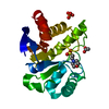
| ||||||||
|---|---|---|---|---|---|---|---|---|---|
| 1 |
| ||||||||
| Unit cell |
| ||||||||
| Details | biological unit is the same as asym. |
-Components
| #1: Protein | Mass: 21729.865 Da / Num. of mol.: 1 / Source method: obtained synthetically |
|---|---|
| #2: Chemical | ChemComp-PO4 /   Mass: 94.971 Da / Num. of mol.: 1 / Source method: obtained synthetically / Formula: PO4 Mass: 94.971 Da / Num. of mol.: 1 / Source method: obtained synthetically / Formula: PO4 |
| #3: Chemical | ChemComp-MES /   Mass: 195.237 Da / Num. of mol.: 1 / Source method: obtained synthetically / Formula: C6H13NO4S / Comment: pH buffer*YM Mass: 195.237 Da / Num. of mol.: 1 / Source method: obtained synthetically / Formula: C6H13NO4S / Comment: pH buffer*YM |
| #4: Chemical | ChemComp-ACY /   Mass: 60.052 Da / Num. of mol.: 1 / Source method: obtained synthetically / Formula: C2H4O2 Mass: 60.052 Da / Num. of mol.: 1 / Source method: obtained synthetically / Formula: C2H4O2 |
| #5: Water | ChemComp-HOH /  Mass: 18.015 Da / Num. of mol.: 75 / Source method: isolated from a natural source / Formula: H2O Mass: 18.015 Da / Num. of mol.: 75 / Source method: isolated from a natural source / Formula: H2O |
-Experimental details
-Experiment
| Experiment | Method: X-RAY DIFFRACTION / Number of used crystals: 1 |
|---|
- Sample preparation
| Crystal | Density Matthews: 2.13 Å3/Da / Density % sol: 42.16 % |
|---|---|
| Crystal grow | Method: microbatch under oil / pH: 6 Details: Protein solution: 100mM NaCl, 5mM DTT, 0.02% NaN3, 10mM Tris-HCl (pH 7.5), Reservoir solution:0.1M Ammonium Sulfate, 0.1M MES, 40% PEG 8000, microbatch under oil |
-Data collection
| Diffraction | Mean temperature: 100 K |
|---|---|
| Diffraction source | Source: SYNCHROTRON / Site: NSLS  / Beamline: X4C / Wavelength: 0.979 Å / Beamline: X4C / Wavelength: 0.979 Å |
| Detector | Type: MAR CCD 165 mm / Detector: CCD / Date: Jun 5, 2012 / Details: mirrors |
| Radiation | Monochromator: Si 111 CHANNEL / Protocol: SINGLE WAVELENGTH / Monochromatic (M) / Laue (L): M / Scattering type: x-ray |
| Radiation wavelength | Wavelength: 0.979 Å / Relative weight: 1 |
| Reflection | Resolution: 2.1→50 Å / Num. obs: 10802 / % possible obs: 95.8 % / Observed criterion σ(I): -3 / Redundancy: 12.7 % / Biso Wilson estimate: 24.2 Å2 / Rmerge(I) obs: 0.081 / Net I/σ(I): 37 |
- Processing
| Software |
| ||||||||||||||||||||||||||||||||||||||||
|---|---|---|---|---|---|---|---|---|---|---|---|---|---|---|---|---|---|---|---|---|---|---|---|---|---|---|---|---|---|---|---|---|---|---|---|---|---|---|---|---|---|
| Refinement | Method to determine structure: BALBES Starting model: 2BFR Resolution: 2.113→41.427 Å / Occupancy max: 1 / Occupancy min: 0.5 / FOM work R set: 0.839 / SU ML: 0.24 / Cross valid method: THROUGHOUT / σ(F): 1.34 / Phase error: 22.42 / Stereochemistry target values: ML
| ||||||||||||||||||||||||||||||||||||||||
| Solvent computation | Shrinkage radii: 0.86 Å / VDW probe radii: 1.1 Å / Solvent model: FLAT BULK SOLVENT MODEL / Bsol: 36.836 Å2 / ksol: 0.378 e/Å3 | ||||||||||||||||||||||||||||||||||||||||
| Displacement parameters | Biso max: 102.19 Å2 / Biso mean: 32.397 Å2 / Biso min: 10.78 Å2
| ||||||||||||||||||||||||||||||||||||||||
| Refinement step | Cycle: LAST / Resolution: 2.113→41.427 Å
| ||||||||||||||||||||||||||||||||||||||||
| Refine LS restraints |
| ||||||||||||||||||||||||||||||||||||||||
| LS refinement shell | Refine-ID: X-RAY DIFFRACTION / Total num. of bins used: 4
| ||||||||||||||||||||||||||||||||||||||||
| Refinement TLS params. | Method: refined / Origin x: -12.6994 Å / Origin y: -15.0508 Å / Origin z: -16.0012 Å
| ||||||||||||||||||||||||||||||||||||||||
| Refinement TLS group |
|
