 Movie
Movie Controller

[English] 日本語
 Yorodumi
- PDB-4gvb: Crystal structure of the virally encoded antifungal protein, KP6,... -
+ Open data

- Basic information
| Entry | Database: PDB / ID: 4gvb | ||||||
|---|---|---|---|---|---|---|---|
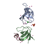
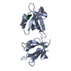
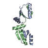
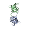
| Title | Crystal structure of the virally encoded antifungal protein, KP6, heterodimer | ||||||
 Components Components |
| ||||||
 Keywords Keywords | TOXIN / Antifungal protein / Secreted | ||||||
| Function / homology | Killer toxin KP6 alpha-subunit / toxin activity / Alpha-Beta Plaits / 2-Layer Sandwich / extracellular region / Alpha Beta / KP6 killer toxin Function and homology information Function and homology information | ||||||
| Biological species |  Ustilago maydis virus P6 Ustilago maydis virus P6 | ||||||
| Method |  X-RAY DIFFRACTION / MOLECULAR REPLACEMENT / Resolution: 1.8 Å X-RAY DIFFRACTION / MOLECULAR REPLACEMENT / Resolution: 1.8 Å | ||||||
 Authors Authors | Smith, T. | ||||||
 Citation Citation | Journal: J.Mol.Biol. / Year: 2013 Title: The Atomic Structure of the Virally Encoded Antifungal Protein, KP6. Authors: Allen, A. / Chatt, E. / Smith, T.J. | ||||||
| History |
|
- Structure visualization
| Structure viewer | Molecule: MolmilJmol/JSmol |
|---|
- Downloads & links
-Download
| PDBx/mmCIF format | 4gvb.cif.gz | 45.3 KB | Display | PDBx/mmCIF format |
|---|---|---|---|---|
| PDB format | pdb4gvb.ent.gz | 32 KB | Display | PDB format |
| PDBx/mmJSON format | 4gvb.json.gz | Tree view | PDBx/mmJSON format | |
| Others |  Other downloads Other downloads |
-Validation report
| Arichive directory | https://data.pdbj.org/pub/pdb/validation_reports/gv/4gvbftp://data.pdbj.org/pub/pdb/validation_reports/gv/4gvb | HTTPS FTP |
|---|
-Related structure data
| Similar structure data |
|---|


-Links
 PDBj
PDBj- Assembly
| Deposited unit | 
| |||||||||
|---|---|---|---|---|---|---|---|---|---|---|
| 1 |
| |||||||||
| Unit cell |
| |||||||||
| Components on special symmetry positions |
|
-Components
| #1: Protein | Mass: 8893.862 Da / Num. of mol.: 1 / Fragment: KP6 alpha subunit / Source method: isolated from a natural source / Source: (natural) Ustilago maydis virus P6 / References: UniProt: P16948 |
|---|---|
| #2: Protein | Mass: 9142.168 Da / Num. of mol.: 1 / Fragment: KP6 beta subunit / Source method: isolated from a natural source / Source: (natural) Ustilago maydis virus P6 / References: UniProt: P16948 |
| #3: Water | ChemComp-HOH /  Mass: 18.015 Da / Num. of mol.: 168 / Source method: isolated from a natural source / Formula: H2O Mass: 18.015 Da / Num. of mol.: 168 / Source method: isolated from a natural source / Formula: H2O |
| Has protein modification | Y |
-Experimental details
-Experiment
| Experiment | Method: X-RAY DIFFRACTION / Number of used crystals: 3 |
|---|
- Sample preparation
| Crystal | Density Matthews: 2.46 Å3/Da / Density % sol: 49.95 % |
|---|---|
| Crystal grow | Temperature: 298 K / Method: vapor diffusion, hanging drop / pH: 8 Details: Reservoir solution contained 50 mM Tris, pH 8.0, 0.7 M ammonium sulfate, and 1 mM sodium azide. The drops were composed of 1:1 ratio of reservoir solution and 8.2 mg/ml KP6, VAPOR DIFFUSION, ...Details: Reservoir solution contained 50 mM Tris, pH 8.0, 0.7 M ammonium sulfate, and 1 mM sodium azide. The drops were composed of 1:1 ratio of reservoir solution and 8.2 mg/ml KP6, VAPOR DIFFUSION, HANGING DROP, temperature 298K |
-Data collection
| Diffraction | Mean temperature: 100 K |
|---|---|
| Diffraction source | Source: ROTATING ANODE / Type: ENRAF-NONIUS FR591 / Wavelength: 1.5418 Å |
| Detector | Type: BRUKER SMART 6000 / Detector: CCD / Date: May 12, 2012 / Details: mirrors |
| Radiation | Monochromator: Osmic (blue) mirror optics / Protocol: SINGLE WAVELENGTH / Monochromatic (M) / Laue (L): M / Scattering type: x-ray |
| Radiation wavelength | Wavelength: 1.5418 Å / Relative weight: 1 |
| Reflection | Resolution: 1.8→39 Å / Num. all: 13869 / Num. obs: 13869 / % possible obs: 93.7 % / Observed criterion σ(F): 1 / Observed criterion σ(I): 1 / Redundancy: 8.2 % |
| Reflection shell | Resolution: 1.8→1.89 Å / % possible all: 90.5 |
- Processing
| Software |
| ||||||||||||||||||||||||||||||||||||||||||
|---|---|---|---|---|---|---|---|---|---|---|---|---|---|---|---|---|---|---|---|---|---|---|---|---|---|---|---|---|---|---|---|---|---|---|---|---|---|---|---|---|---|---|---|
| Refinement | Method to determine structure: MOLECULAR REPLACEMENT / Resolution: 1.8→38.468 Å / SU ML: 0.23 / σ(F): 0.08 / Phase error: 24.77 / Stereochemistry target values: ML
| ||||||||||||||||||||||||||||||||||||||||||
| Solvent computation | Shrinkage radii: 0.9 Å / VDW probe radii: 1.11 Å / Solvent model: FLAT BULK SOLVENT MODEL / Bsol: 50.721 Å2 / ksol: 0.357 e/Å3 | ||||||||||||||||||||||||||||||||||||||||||
| Displacement parameters |
| ||||||||||||||||||||||||||||||||||||||||||
| Refinement step | Cycle: LAST / Resolution: 1.8→38.468 Å
| ||||||||||||||||||||||||||||||||||||||||||
| Refine LS restraints |
| ||||||||||||||||||||||||||||||||||||||||||
| LS refinement shell |
|
