- PDB-4gsq: Structural basis for the inhibition of Mycobacterium tuberculosis... -
+
Open data
ID or keywords:
Loading...
-
Basic information
Entry
Database: PDB / ID: 4gsq
Title
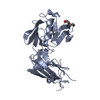
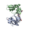
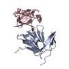
Structural basis for the inhibition of Mycobacterium tuberculosis L,D-transpeptidase by meropenem, a drug effective against extensively drug-resistant strains
Components
Probable conserved lipoprotein LPPS
Keywords
TRANSFERASE / L / D-transpeptidase
Function / homology
Function and homology information
peptidoglycan-based cell wall biogenesis / peptidoglycan-protein cross-linking / peptidoglycan metabolic process / peptidoglycan L,D-transpeptidase activity / Transferases; Acyltransferases; Aminoacyltransferases / acyltransferase activity / peptidoglycan-based cell wall / cell wall organization / regulation of cell shape / extracellular region ...peptidoglycan-based cell wall biogenesis / peptidoglycan-protein cross-linking / peptidoglycan metabolic process / peptidoglycan L,D-transpeptidase activity / Transferases; Acyltransferases; Aminoacyltransferases / acyltransferase activity / peptidoglycan-based cell wall / cell wall organization / regulation of cell shape / extracellular region / metal ion binding / plasma membrane Similarity search - Function
Method to determine structure: SAD / Resolution: 1.8→20 Å / Cor.coef. Fo:Fc: 0.952 / Cor.coef. Fo:Fc free: 0.93 / SU B: 2.298 / SU ML: 0.073 / Cross valid method: THROUGHOUT / ESU R: 0.118 / ESU R Free: 0.118 / Stereochemistry target values: MAXIMUM LIKELIHOOD / Details: HYDROGENS HAVE BEEN USED IF PRESENT IN THE INPUT
Rfactor
Num. reflection
% reflection
Selection details
Rfree
0.23346
1489
5.1 %
RANDOM
Rwork
0.19469
-
-
-
all
0.19663
28536
-
-
obs
0.19663
27937
99.04 %
-
Solvent computation
Ion probe radii: 0.8 Å / Shrinkage radii: 0.8 Å / VDW probe radii: 1.2 Å / Solvent model: MASK
Displacement parameters
Biso mean: 24.367 Å2
Baniso -1
Baniso -2
Baniso -3
1-
-1.52 Å2
0 Å2
0.2 Å2
2-
-
-0.38 Å2
-0 Å2
3-
-
-
1.93 Å2
Refinement step
Cycle: LAST / Resolution: 1.8→20 Å
Protein
Nucleic acid
Ligand
Solvent
Total
Num. atoms
1889
0
7
206
2102
Refine LS restraints
Refine-ID
Type
Dev ideal
Dev ideal target
Number
X-RAY DIFFRACTION
r_bond_refined_d
0.009
0.02
1945
X-RAY DIFFRACTION
r_angle_refined_deg
1.292
1.913
2655
X-RAY DIFFRACTION
r_dihedral_angle_1_deg
5.568
5
243
X-RAY DIFFRACTION
r_dihedral_angle_2_deg
29.684
24.396
91
X-RAY DIFFRACTION
r_dihedral_angle_3_deg
13.234
15
285
X-RAY DIFFRACTION
r_dihedral_angle_4_deg
12.278
15
10
X-RAY DIFFRACTION
r_chiral_restr
0.088
0.2
288
X-RAY DIFFRACTION
r_gen_planes_refined
0.006
0.021
1517
LS refinement shell
Resolution: 1.802→1.849 Å / Total num. of bins used: 20
Rfactor
Num. reflection
% reflection
Rfree
0.307
106
-
Rwork
0.247
1946
-
obs
-
-
96.7 %
+
About Yorodumi
-
News
-
Feb 9, 2022. New format data for meta-information of EMDB entries
New format data for meta-information of EMDB entries
Version 3 of the EMDB header file is now the official format.
The previous official version 1.9 will be removed from the archive.
In the structure databanks used in Yorodumi, some data are registered as the other names, "COVID-19 virus" and "2019-nCoV". Here are the details of the virus and the list of structure data.
Jan 31, 2019. EMDB accession codes are about to change! (news from PDBe EMDB page)
EMDB accession codes are about to change! (news from PDBe EMDB page)
The allocation of 4 digits for EMDB accession codes will soon come to an end. Whilst these codes will remain in use, new EMDB accession codes will include an additional digit and will expand incrementally as the available range of codes is exhausted. The current 4-digit format prefixed with “EMD-” (i.e. EMD-XXXX) will advance to a 5-digit format (i.e. EMD-XXXXX), and so on. It is currently estimated that the 4-digit codes will be depleted around Spring 2019, at which point the 5-digit format will come into force.
The EM Navigator/Yorodumi systems omit the EMD- prefix.
Related info.:Q: What is EMD? / ID/Accession-code notation in Yorodumi/EM Navigator
Yorodumi is a browser for structure data from EMDB, PDB, SASBDB, etc.
This page is also the successor to EM Navigator detail page, and also detail information page/front-end page for Omokage search.
The word "yorodu" (or yorozu) is an old Japanese word meaning "ten thousand". "mi" (miru) is to see.
Related info.:EMDB / PDB / SASBDB / Comparison of 3 databanks / Yorodumi Search / Aug 31, 2016. New EM Navigator & Yorodumi / Yorodumi Papers / Jmol/JSmol / Function and homology information / Changes in new EM Navigator and Yorodumi
 Movie
Movie Controller

 Yorodumi
 Open data
 Basic information
 Components
 Keywords
 Function and homology information
 Mycobacterium tuberculosis (bacteria)
 X-RAY DIFFRACTION /
  Authors
 Citation
 Structure visualization
 Downloads & links
 Other downloads


 PDBj
PDBj


 Assembly
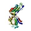

 Mass: 40.078 Da / Num. of mol.: 1 / Source method: obtained synthetically / Formula: Ca
 Mass: 92.094 Da / Num. of mol.: 1 / Source method: obtained synthetically / Formula: C3H8O3
 Mass: 18.015 Da / Num. of mol.: 206 / Source method: isolated from a natural source / Formula: H2O
 Sample preparation
 / Beamline: AR-NE3A / Wavelength: 1 Å
 Processing