 Movie
Movie Controller

[English] 日本語
 Yorodumi
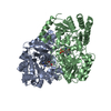
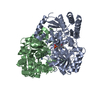
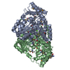
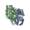
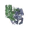
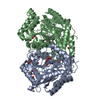
- PDB-4gsa: CRYSTAL STRUCTURE OF GLUTAMATE-1-SEMIALDEHYDE AMINOMUTASE (AMINOT... -
+ Open data

- Basic information
| Entry | Database: PDB / ID: 4gsa | ||||||
|---|---|---|---|---|---|---|---|
| Title | CRYSTAL STRUCTURE OF GLUTAMATE-1-SEMIALDEHYDE AMINOMUTASE (AMINOTRANSFERASE) REDUCED WITH CYANOBOROHYDRATE | ||||||
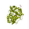
 Components Components | GLUTAMATE SEMIALDEHYDE AMINOTRANSFERASE | ||||||
 Keywords Keywords | CHLOROPHYLL BIOSYNTHESIS / PYRIDOXAL-5'-PHOSPHATE / PYRIDOXAMINE-5'-PHOSPHATE / ASYMMETRIC DIMER / REDUCED FORM | ||||||
| Function / homology |  Function and homology information Function and homology informationglutamate-1-semialdehyde 2,1-aminomutase / glutamate-1-semialdehyde 2,1-aminomutase activity / chlorophyll biosynthetic process / : / transaminase activity / pyridoxal phosphate binding / cytoplasm Similarity search - Function | ||||||
| Biological species |  Synechococcus sp. (bacteria) Synechococcus sp. (bacteria) | ||||||
| Method |  X-RAY DIFFRACTION / Resolution: 2.5 Å X-RAY DIFFRACTION / Resolution: 2.5 Å | ||||||
 Authors Authors | Hennig, M. / Jansonius, J.N. | ||||||
 Citation Citation | Journal: Proc.Natl.Acad.Sci.USA / Year: 1997 Title: Crystal structure of glutamate-1-semialdehyde aminomutase: an alpha2-dimeric vitamin B6-dependent enzyme with asymmetry in structure and active site reactivity. Authors: Hennig, M. / Grimm, B. / Contestabile, R. / John, R.A. / Jansonius, J.N. #1: Journal: J.Mol.Biol. / Year: 1994Title: Crystallization and Preliminary X-Ray Analysis of Wild-Type and K272A Mutant Glutamate 1-Semialdehyde Aminotransferase from Synechococcus Authors: Hennig, M. / Grimm, B. / Jenny, M. / Muller, R. / Jansonius, J.N. | ||||||
| History |
|
- Structure visualization
| Structure viewer | Molecule: MolmilJmol/JSmol |
|---|
- Downloads & links
-Download
| PDBx/mmCIF format | 4gsa.cif.gz | 172.8 KB | Display | PDBx/mmCIF format |
|---|---|---|---|---|
| PDB format | pdb4gsa.ent.gz | 136.8 KB | Display | PDB format |
| PDBx/mmJSON format | 4gsa.json.gz | Tree view | PDBx/mmJSON format | |
| Others |  Other downloads Other downloads |
-Validation report
| Arichive directory | https://data.pdbj.org/pub/pdb/validation_reports/gs/4gsaftp://data.pdbj.org/pub/pdb/validation_reports/gs/4gsa | HTTPS FTP |
|---|
-Related structure data


-Links
 PDBj
PDBj- Assembly
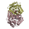
| Deposited unit | 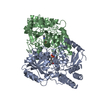
| ||||||||
|---|---|---|---|---|---|---|---|---|---|
| 1 |
| ||||||||
| Unit cell |
|
-Components
| #1: Protein | Mass: 46059.523 Da / Num. of mol.: 2 Source method: isolated from a genetically manipulated source Source: (gene. exp.) Synechococcus sp. (bacteria) / Strain: GR6 / Production host: References: UniProt: P24630, glutamate-1-semialdehyde 2,1-aminomutase #2: Chemical |   Mass: 247.142 Da / Num. of mol.: 2 / Source method: obtained synthetically / Formula: C8H10NO6P Mass: 247.142 Da / Num. of mol.: 2 / Source method: obtained synthetically / Formula: C8H10NO6P#3: Water | ChemComp-HOH / |  Mass: 18.015 Da / Num. of mol.: 190 / Source method: isolated from a natural source / Formula: H2O Mass: 18.015 Da / Num. of mol.: 190 / Source method: isolated from a natural source / Formula: H2O |
|---|
-Experimental details
-Experiment
| Experiment | Method: X-RAY DIFFRACTION / Number of used crystals: 1 |
|---|
- Sample preparation
| Crystal | Density Matthews: 2.4 Å3/Da / Density % sol: 50 % Description: COFACTORS AND WATER MOLECULES WERE REJECTED FROM THE MODEL. | |||||||||||||||||||||||||||||||||||
|---|---|---|---|---|---|---|---|---|---|---|---|---|---|---|---|---|---|---|---|---|---|---|---|---|---|---|---|---|---|---|---|---|---|---|---|---|
| Crystal grow | pH: 7 Details: 50MM NA-CACODYLATE BUFFER PH 7.0, 200 MM MG-ACETATE, 19.5% PEG 10,000 | |||||||||||||||||||||||||||||||||||
| Crystal grow | *PLUS Temperature: 4 ℃ / Method: vapor diffusion, sitting drop / Details: Hennig, M., (1994) J.Mol.Biol., 242, 591. | |||||||||||||||||||||||||||||||||||
| Components of the solutions | *PLUS
|
-Data collection
| Diffraction | Mean temperature: 273 K |
|---|---|
| Diffraction source | Source: ROTATING ANODE / Type: ELLIOTT GX-21 / Wavelength: 1.5418 |
| Detector | Type: MARRESEARCH / Detector: IMAGE PLATE / Date: Apr 7, 1995 |
| Radiation | Monochromator: GRAPHITE(002) / Monochromatic (M) / Laue (L): M / Scattering type: x-ray |
| Radiation wavelength | Wavelength: 1.5418 Å / Relative weight: 1 |
| Reflection | Resolution: 2.5→15 Å / Num. obs: 31141 / % possible obs: 96.8 % / Redundancy: 2.9 % / Biso Wilson estimate: 30 Å2 / Rsym value: 0.073 / Net I/σ(I): 9.7 |
| Reflection shell | Resolution: 2.5→2.63 Å / Redundancy: 2.8 % / Mean I/σ(I) obs: 2 / Rsym value: 37.2 / % possible all: 91.7 |
| Reflection | *PLUS Num. measured all: 91742 / Rmerge(I) obs: 0.073 |
- Processing
| Software |
| ||||||||||||||||||||||||||||||||||||||||||||||||||||||||||||
|---|---|---|---|---|---|---|---|---|---|---|---|---|---|---|---|---|---|---|---|---|---|---|---|---|---|---|---|---|---|---|---|---|---|---|---|---|---|---|---|---|---|---|---|---|---|---|---|---|---|---|---|---|---|---|---|---|---|---|---|---|---|
| Refinement | Starting model: GSAT WILD-TYPE STRUCTURE Resolution: 2.5→15 Å
| ||||||||||||||||||||||||||||||||||||||||||||||||||||||||||||
| Displacement parameters | Biso mean: 28.4 Å2 | ||||||||||||||||||||||||||||||||||||||||||||||||||||||||||||
| Refinement step | Cycle: LAST / Resolution: 2.5→15 Å
| ||||||||||||||||||||||||||||||||||||||||||||||||||||||||||||
| Refine LS restraints |
| ||||||||||||||||||||||||||||||||||||||||||||||||||||||||||||
| LS refinement shell | Resolution: 2.5→2.54 Å / Total num. of bins used: 20
|
