 Movie
Movie Controller


+ Open data

- Basic information
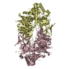
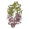
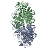
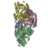
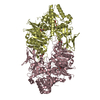
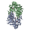
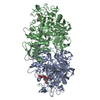
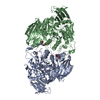
| Entry | Database: PDB / ID: 4gq2 | ||||||
|---|---|---|---|---|---|---|---|
| Title | S. pombe Nup120-Nup37 complex | ||||||
 Components Components |
| ||||||
 Keywords Keywords | TRANSPORT PROTEIN / Beta propeller alpha helical / component of nuclear pore complex | ||||||
| Function / homology |  Function and homology information Function and homology informationTransport of the SLBP independent Mature mRNA / Transport of Mature mRNA Derived from an Intronless Transcript / Transport of Mature mRNA derived from an Intron-Containing Transcript / Regulation of Glucokinase by Glucokinase Regulatory Protein / Regulation of HSF1-mediated heat shock response / SUMOylation of SUMOylation proteins / SUMOylation of RNA binding proteins / Postmitotic nuclear pore complex (NPC) reformation / SUMOylation of chromatin organization proteins / Transcriptional regulation by small RNAs ...Transport of the SLBP independent Mature mRNA / Transport of Mature mRNA Derived from an Intronless Transcript / Transport of Mature mRNA derived from an Intron-Containing Transcript / Regulation of Glucokinase by Glucokinase Regulatory Protein / Regulation of HSF1-mediated heat shock response / SUMOylation of SUMOylation proteins / SUMOylation of RNA binding proteins / Postmitotic nuclear pore complex (NPC) reformation / SUMOylation of chromatin organization proteins / Transcriptional regulation by small RNAs / nuclear pore outer ring / structural constituent of nuclear pore / nucleocytoplasmic transport / poly(A)+ mRNA export from nucleus / nuclear pore / nuclear periphery / nuclear envelope / nucleus / cytosol Similarity search - Function | ||||||
| Biological species |  | ||||||
| Method |  X-RAY DIFFRACTION / SYNCHROTRON / molecular replacement-SAD / Resolution: 2.4 Å X-RAY DIFFRACTION / SYNCHROTRON / molecular replacement-SAD / Resolution: 2.4 Å | ||||||
 Authors Authors | Liu, X. / Mitchell, J. / Wozniak, R. / Blobel, G. / Fan, J. | ||||||
 Citation Citation | Journal: Proc.Natl.Acad.Sci.USA / Year: 2012 Title: Structural evolution of the membrane-coating module of the nuclear pore complex. Authors: Liu, X. / Mitchell, J.M. / Wozniak, R.W. / Blobel, G. / Fan, J. | ||||||
| History |
|
- Structure visualization
| Structure viewer | Molecule: MolmilJmol/JSmol |
|---|
- Downloads & links
-Download
| PDBx/mmCIF format | 4gq2.cif.gz | 267.2 KB | Display | PDBx/mmCIF format |
|---|---|---|---|---|
| PDB format | pdb4gq2.ent.gz | 211.7 KB | Display | PDB format |
| PDBx/mmJSON format | 4gq2.json.gz | Tree view | PDBx/mmJSON format | |
| Others |  Other downloads Other downloads |
-Validation report
| Arichive directory | https://data.pdbj.org/pub/pdb/validation_reports/gq/4gq2ftp://data.pdbj.org/pub/pdb/validation_reports/gq/4gq2 | HTTPS FTP |
|---|
-Related structure data
| Related structure data |  4gq1SC S: Starting model for refinement C: citing same article ( |
|---|---|
| Similar structure data |


-Links
 PDBj
PDBj

- Assembly
| Deposited unit | 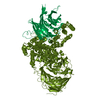
| ||||||||
|---|---|---|---|---|---|---|---|---|---|
| 1 |
| ||||||||
| Unit cell |
|
-Components
| #1: Protein | Mass: 108344.375 Da / Num. of mol.: 1 Source method: isolated from a genetically manipulated source Source: (gene. exp.) Gene: nup120, SPBC3B9.16c / Plasmid: pFastBac / Production host:   Spodoptera frugiperda (fall armyworm) / Strain (production host): Hi5 / References: UniProt: O43044 Spodoptera frugiperda (fall armyworm) / Strain (production host): Hi5 / References: UniProt: O43044 |
|---|---|
| #2: Protein | Mass: 42951.199 Da / Num. of mol.: 1 Source method: isolated from a genetically manipulated source Source: (gene. exp.) Gene: Nup37, SPAC4F10.18 / Plasmid: pFastBac / Production host: Spodoptera frugiperda (fall armyworm) / Strain (production host): Hi5 / References: UniProt: O36030 |
| #3: Water | ChemComp-HOH /  Mass: 18.015 Da / Num. of mol.: 122 / Source method: isolated from a natural source / Formula: H2O Mass: 18.015 Da / Num. of mol.: 122 / Source method: isolated from a natural source / Formula: H2O |
-Experimental details
-Experiment
| Experiment | Method: X-RAY DIFFRACTION / Number of used crystals: 1 |
|---|
- Sample preparation
| Crystal | Density Matthews: 2.49 Å3/Da / Density % sol: 50.57 % |
|---|---|
| Crystal grow | Temperature: 303 K / Method: evaporation / pH: 5.5 Details: 10% PEG3350, 0.1 M Bis-Tris, pH 5.5, 0.2 M ammonium acetate, EVAPORATION, temperature 303K |
-Data collection
| Diffraction | Mean temperature: 100 K |
|---|---|
| Diffraction source | Source: SYNCHROTRON / Site: NSLS  / Beamline: X29A / Wavelength: 1.075 Å / Beamline: X29A / Wavelength: 1.075 Å |
| Detector | Type: ADSC QUANTUM 315r / Detector: CCD / Date: Apr 27, 2012 |
| Radiation | Monochromator: Rosenbaum-Rock double crystal sagittal focusing Protocol: SINGLE WAVELENGTH / Monochromatic (M) / Laue (L): M / Scattering type: x-ray |
| Radiation wavelength | Wavelength: 1.075 Å / Relative weight: 1 |
| Reflection | Resolution: 2.4→35 Å / Num. all: 56356 / Num. obs: 56356 / % possible obs: 100 % / Observed criterion σ(F): 0 / Observed criterion σ(I): 0 / Redundancy: 7.2 % / Biso Wilson estimate: 40 Å2 / Rmerge(I) obs: 0.07 / Rsym value: 0.07 / Net I/σ(I): 34 |
| Reflection shell | Resolution: 2.4→2.46 Å / Redundancy: 7.6 % / Rmerge(I) obs: 0.91 / Mean I/σ(I) obs: 2.6 / Rsym value: 0.91 / % possible all: 100 |
- Processing
| Software |
| ||||||||||||||||||||||||||||||||||||||||||||||||||||||||||||||||||||||||||||||||||||||||||||||||||||||||||||||||||||||||||||||||||||||||||||||||||||||||||||||||||||||||||
|---|---|---|---|---|---|---|---|---|---|---|---|---|---|---|---|---|---|---|---|---|---|---|---|---|---|---|---|---|---|---|---|---|---|---|---|---|---|---|---|---|---|---|---|---|---|---|---|---|---|---|---|---|---|---|---|---|---|---|---|---|---|---|---|---|---|---|---|---|---|---|---|---|---|---|---|---|---|---|---|---|---|---|---|---|---|---|---|---|---|---|---|---|---|---|---|---|---|---|---|---|---|---|---|---|---|---|---|---|---|---|---|---|---|---|---|---|---|---|---|---|---|---|---|---|---|---|---|---|---|---|---|---|---|---|---|---|---|---|---|---|---|---|---|---|---|---|---|---|---|---|---|---|---|---|---|---|---|---|---|---|---|---|---|---|---|---|---|---|---|---|---|
| Refinement | Method to determine structure: molecular replacement-SAD Starting model: PDB ENTRY 4GQ1 Resolution: 2.4→35 Å / Cor.coef. Fo:Fc: 0.952 / Cor.coef. Fo:Fc free: 0.929 / SU B: 9.658 / SU ML: 0.22 / Cross valid method: THROUGHOUT / ESU R: 0.417 / ESU R Free: 0.269 / Stereochemistry target values: MAXIMUM LIKELIHOOD
| ||||||||||||||||||||||||||||||||||||||||||||||||||||||||||||||||||||||||||||||||||||||||||||||||||||||||||||||||||||||||||||||||||||||||||||||||||||||||||||||||||||||||||
| Solvent computation | Ion probe radii: 0.8 Å / Shrinkage radii: 0.8 Å / VDW probe radii: 1.2 Å / Solvent model: BABINET MODEL WITH MASK | ||||||||||||||||||||||||||||||||||||||||||||||||||||||||||||||||||||||||||||||||||||||||||||||||||||||||||||||||||||||||||||||||||||||||||||||||||||||||||||||||||||||||||
| Displacement parameters | Biso mean: 62.694 Å2
| ||||||||||||||||||||||||||||||||||||||||||||||||||||||||||||||||||||||||||||||||||||||||||||||||||||||||||||||||||||||||||||||||||||||||||||||||||||||||||||||||||||||||||
| Refinement step | Cycle: LAST / Resolution: 2.4→35 Å
| ||||||||||||||||||||||||||||||||||||||||||||||||||||||||||||||||||||||||||||||||||||||||||||||||||||||||||||||||||||||||||||||||||||||||||||||||||||||||||||||||||||||||||
| Refine LS restraints |
| ||||||||||||||||||||||||||||||||||||||||||||||||||||||||||||||||||||||||||||||||||||||||||||||||||||||||||||||||||||||||||||||||||||||||||||||||||||||||||||||||||||||||||
| LS refinement shell | Resolution: 2.4→2.462 Å / Total num. of bins used: 20
|
