 Movie
Movie Controller

[English] 日本語
 Yorodumi
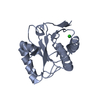
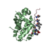
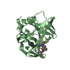
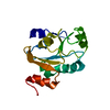
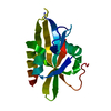
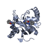
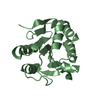
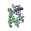
- PDB-4gkf: Crystal structure and characterization of Cmr5 protein from Pyroc... -
+ Open data

- Basic information
| Entry | Database: PDB / ID: 4gkf | ||||||
|---|---|---|---|---|---|---|---|
| Title | Crystal structure and characterization of Cmr5 protein from Pyrococcus furiosus | ||||||
 Components Components | CRISPR system Cmr subunit Cmr5 | ||||||
 Keywords Keywords | UNKNOWN FUNCTION / Crispr | ||||||
| Function / homology |  Function and homology information Function and homology information | ||||||
| Biological species |   Pyrococcus furiosus (archaea) Pyrococcus furiosus (archaea) | ||||||
| Method |  X-RAY DIFFRACTION / SYNCHROTRON / MOLECULAR REPLACEMENT / Resolution: 2.1 Å X-RAY DIFFRACTION / SYNCHROTRON / MOLECULAR REPLACEMENT / Resolution: 2.1 Å | ||||||
 Authors Authors | Park, J. / Sun, J. / Park, S. / Hwang, H. / Park, M. / Shin, M.S. | ||||||
 Citation Citation | Journal: Febs Lett. / Year: 2013 Title: Crystal structure of Cmr5 from Pyrococcus furiosus and its functional implications Authors: Park, J.H. / Sun, J. / Park, S.Y. / Hwang, H.J. / Park, M.Y. / Shin, M. / Kim, J.S. | ||||||
| History |
|
- Structure visualization
| Structure viewer | Molecule: MolmilJmol/JSmol |
|---|
- Downloads & links
-Download
| PDBx/mmCIF format | 4gkf.cif.gz | 74.6 KB | Display | PDBx/mmCIF format |
|---|---|---|---|---|
| PDB format | pdb4gkf.ent.gz | 56 KB | Display | PDB format |
| PDBx/mmJSON format | 4gkf.json.gz | Tree view | PDBx/mmJSON format | |
| Others |  Other downloads Other downloads |
-Validation report
| Arichive directory | https://data.pdbj.org/pub/pdb/validation_reports/gk/4gkfftp://data.pdbj.org/pub/pdb/validation_reports/gk/4gkf | HTTPS FTP |
|---|
-Related structure data
| Related structure data |  2oebS S: Starting model for refinement |
|---|---|
| Similar structure data |
-Links
 PDBj
PDBj
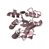
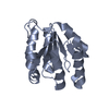
- Assembly
| Deposited unit | 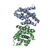
| ||||||||
|---|---|---|---|---|---|---|---|---|---|
| 1 | 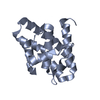
| ||||||||
| 2 | 
| ||||||||
| Unit cell |
|
-Components
| #1: Protein | Mass: 19716.793 Da / Num. of mol.: 2 Source method: isolated from a genetically manipulated source Source: (gene. exp.) Pyrococcus furiosus (archaea) / Strain: ATCC 43587 / DSM 3638 / JCM 8422 / Vc1 / Gene: cmr5, PF1125 / Production host:  #2: Water | ChemComp-HOH / |  Mass: 18.015 Da / Num. of mol.: 184 / Source method: isolated from a natural source / Formula: H2O Mass: 18.015 Da / Num. of mol.: 184 / Source method: isolated from a natural source / Formula: H2O |
|---|
-Experimental details
-Experiment
| Experiment | Method: X-RAY DIFFRACTION / Number of used crystals: 1 |
|---|
- Sample preparation
| Crystal | Density Matthews: 2.18 Å3/Da / Density % sol: 43.49 % |
|---|---|
| Crystal grow | Temperature: 295 K / Method: vapor diffusion, hanging drop / pH: 7.5 Details: 20 %(w/v) Polyethylene Glycol 3350, 0.1M Bis-Tris HCl, 0.2M Magnesium Chloride, pH 7.5, VAPOR DIFFUSION, HANGING DROP, temperature 295K |
-Data collection
| Diffraction | Mean temperature: 100 K |
|---|---|
| Diffraction source | Source: SYNCHROTRON / Site: SSRF  / Beamline: BL17U / Wavelength: 0.9762 Å / Beamline: BL17U / Wavelength: 0.9762 Å |
| Detector | Type: ADSC QUANTUM 315 / Detector: CCD / Date: May 12, 2011 |
| Radiation | Monochromator: Graphite / Protocol: SINGLE WAVELENGTH / Monochromatic (M) / Laue (L): M / Scattering type: x-ray |
| Radiation wavelength | Wavelength: 0.9762 Å / Relative weight: 1 |
| Reflection | Resolution: 2.1→50 Å / Num. obs: 23080 / % possible obs: 93.2 % / Observed criterion σ(F): -3 / Observed criterion σ(I): -3 |
| Reflection shell | Resolution: 2.1→2.15 Å / % possible all: 91 |
- Processing
| Software |
| ||||||||||||||||||||
|---|---|---|---|---|---|---|---|---|---|---|---|---|---|---|---|---|---|---|---|---|---|
| Refinement | Method to determine structure: MOLECULAR REPLACEMENT Starting model: 2oeb Resolution: 2.1→49.06 Å / σ(F): 0 / Stereochemistry target values: Engh & Huber
| ||||||||||||||||||||
| Refinement step | Cycle: LAST / Resolution: 2.1→49.06 Å
| ||||||||||||||||||||
| Refine LS restraints |
|
