 Movie
Movie Controller


+ Open data

- Basic information
| Entry | Database: PDB / ID: 4gk0 | ||||||
|---|---|---|---|---|---|---|---|
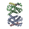
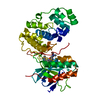
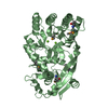
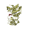
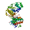
| Title | Crystal structure of human Rev3-Rev7-Rev1 complex | ||||||
 Components Components |
| ||||||
 Keywords Keywords | TRANSFERASE / translesion polymerases complex / four-helix bundle / beta-hairpin domain / anti-parallel sheets / translesion DNA synthesis / polymerase switch / none | ||||||
| Function / homology |  Function and homology information Function and homology informationsomatic diversification of immunoglobulins involved in immune response / DNA damage response, signal transduction resulting in transcription / deoxycytidyl transferase activity / negative regulation of ubiquitin protein ligase activity / zeta DNA polymerase complex / DNA-(abasic site) binding / positive regulation of isotype switching / : / negative regulation of cell-cell adhesion mediated by cadherin / JUN kinase binding ...somatic diversification of immunoglobulins involved in immune response / DNA damage response, signal transduction resulting in transcription / deoxycytidyl transferase activity / negative regulation of ubiquitin protein ligase activity / zeta DNA polymerase complex / DNA-(abasic site) binding / positive regulation of isotype switching / : / negative regulation of cell-cell adhesion mediated by cadherin / JUN kinase binding / error-free translesion synthesis / negative regulation of epithelial to mesenchymal transition / DNA biosynthetic process / positive regulation of double-strand break repair via nonhomologous end joining / mitotic spindle assembly checkpoint signaling / DNA synthesis involved in DNA repair / telomere maintenance in response to DNA damage / nuclear replication fork / positive regulation of peptidyl-serine phosphorylation / error-prone translesion synthesis / negative regulation of double-strand break repair via homologous recombination / somatic hypermutation of immunoglobulin genes / site of DNA damage / translesion synthesis / Translesion synthesis by REV1 / Translesion synthesis by POLK / actin filament organization / Translesion synthesis by POLI / ubiquitin binding / Termination of translesion DNA synthesis / regulation of cell growth / negative regulation of canonical Wnt signaling pathway / negative regulation of protein catabolic process / DNA-templated DNA replication / double-strand break repair via homologous recombination / spindle / Transferases; Transferring phosphorus-containing groups; Nucleotidyltransferases / transcription corepressor activity / double-strand break repair / chromosome / site of double-strand break / 4 iron, 4 sulfur cluster binding / DNA-directed DNA polymerase / damaged DNA binding / RNA polymerase II-specific DNA-binding transcription factor binding / DNA-directed DNA polymerase activity / protein-macromolecule adaptor activity / cell division / nucleotide binding / DNA repair / positive regulation of DNA-templated transcription / chromatin / nucleolus / negative regulation of transcription by RNA polymerase II / DNA binding / zinc ion binding / nucleoplasm / metal ion binding / nucleus / cytoplasm Similarity search - Function | ||||||
| Biological species |  Homo sapiens (human) Homo sapiens (human) | ||||||
| Method |  X-RAY DIFFRACTION / SYNCHROTRON / MOLECULAR REPLACEMENT / Resolution: 2.7 Å X-RAY DIFFRACTION / SYNCHROTRON / MOLECULAR REPLACEMENT / Resolution: 2.7 Å | ||||||
 Authors Authors | Tao, J. / Min, X. / Wei, X. | ||||||
 Citation Citation | Journal: Protein Cell / Year: 2012 Title: Structural insights into the assembly of human translesion polymerase complexes Authors: Xie, W. / Yang, X. / Xu, M. / Jiang, T. | ||||||
| History |
|
- Structure visualization
| Structure viewer | Molecule: MolmilJmol/JSmol |
|---|
- Downloads & links
-Download
| PDBx/mmCIF format | 4gk0.cif.gz | 136.6 KB | Display | PDBx/mmCIF format |
|---|---|---|---|---|
| PDB format | pdb4gk0.ent.gz | 105.6 KB | Display | PDB format |
| PDBx/mmJSON format | 4gk0.json.gz | Tree view | PDBx/mmJSON format | |
| Others |  Other downloads Other downloads |
-Validation report
| Arichive directory | https://data.pdbj.org/pub/pdb/validation_reports/gk/4gk0ftp://data.pdbj.org/pub/pdb/validation_reports/gk/4gk0 | HTTPS FTP |
|---|
-Related structure data
| Related structure data | 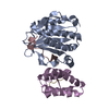 4gk5C 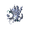 3abdS C: citing same article ( S: Starting model for refinement |
|---|---|
| Similar structure data |


-Links
 PDBj
PDBj


- Assembly
| Deposited unit | 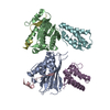
| ||||||||
|---|---|---|---|---|---|---|---|---|---|
| 1 | 
| ||||||||
| 2 | 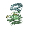
| ||||||||
| Unit cell |
|
-Components
| #1: Protein | Mass: 26960.033 Da / Num. of mol.: 2 / Mutation: R124A Source method: isolated from a genetically manipulated source Source: (gene. exp.) Homo sapiens (human) / Gene: MAD2L2, MAD2B, REV7 / Plasmid: pETDuet-1 / Production host:  #2: Protein | Mass: 5632.319 Da / Num. of mol.: 2 / Fragment: Rev7-binding domain, UNP residues 1847-1898 Source method: isolated from a genetically manipulated source Source: (gene. exp.) Homo sapiens (human) / Gene: REV3L, POLZ, REV3 / Plasmid: pETDuet-1 / Production host: #3: Protein | Mass: 15179.386 Da / Num. of mol.: 2 / Fragment: C-terminal domain, UNP residues 1117-1251 Source method: isolated from a genetically manipulated source Source: (gene. exp.) Homo sapiens (human) / Gene: REV1, REV1L / Plasmid: pETDuet-1 / Production host: References: UniProt: Q9UBZ9, Transferases; Transferring phosphorus-containing groups; Nucleotidyltransferases |
|---|
-Experimental details
-Experiment
| Experiment | Method: X-RAY DIFFRACTION / Number of used crystals: 1 |
|---|
- Sample preparation
| Crystal | Density Matthews: 2.42 Å3/Da / Density % sol: 49.2 % |
|---|---|
| Crystal grow | Temperature: 289 K / Method: vapor diffusion, hanging drop / pH: 5.8 Details: 0.1M sodium citrate, 1.95M sodium formate, 20mM DTT , pH 5.8, VAPOR DIFFUSION, HANGING DROP, temperature 289K |
-Data collection
| Diffraction | Mean temperature: 100 K |
|---|---|
| Diffraction source | Source: SYNCHROTRON / Site: SSRF  / Beamline: BL17U / Wavelength: 0.9791 Å / Beamline: BL17U / Wavelength: 0.9791 Å |
| Detector | Type: ADSC QUANTUM 315r / Detector: CCD / Date: Jun 1, 2011 |
| Radiation | Monochromator: GRAPHITE / Protocol: SINGLE WAVELENGTH / Monochromatic (M) / Laue (L): M / Scattering type: x-ray |
| Radiation wavelength | Wavelength: 0.9791 Å / Relative weight: 1 |
| Reflection | Resolution: 2.7→20 Å / Num. all: 23773 / Num. obs: 23773 / % possible obs: 95.9 % / Observed criterion σ(F): 0 / Observed criterion σ(I): 0 / Redundancy: 3.3 % / Rmerge(I) obs: 0.063 / Net I/σ(I): 22.3 |
| Reflection shell | Resolution: 2.7→2.8 Å / Redundancy: 3.2 % / Rmerge(I) obs: 0.383 / Mean I/σ(I) obs: 3.3 / Num. unique all: 2511 / % possible all: 97.5 |
- Processing
| Software |
| ||||||||||||||||||||||||||||||||||||||||||||||||||||||||||||
|---|---|---|---|---|---|---|---|---|---|---|---|---|---|---|---|---|---|---|---|---|---|---|---|---|---|---|---|---|---|---|---|---|---|---|---|---|---|---|---|---|---|---|---|---|---|---|---|---|---|---|---|---|---|---|---|---|---|---|---|---|---|
| Refinement | Method to determine structure: MOLECULAR REPLACEMENT Starting model: 3ABD Resolution: 2.7→20 Å / Cor.coef. Fo:Fc: 0.938 / Cor.coef. Fo:Fc free: 0.905 / Cross valid method: THROUGHOUT / σ(F): 0 / σ(I): 0 / ESU R: 0.457 / ESU R Free: 0.349 / Stereochemistry target values: MAXIMUM LIKELIHOOD / Details: HYDROGENS HAVE BEEN ADDED IN THE RIDING POSITIONS
| ||||||||||||||||||||||||||||||||||||||||||||||||||||||||||||
| Solvent computation | Ion probe radii: 0.8 Å / Shrinkage radii: 0.8 Å / VDW probe radii: 1.2 Å / Solvent model: MASK | ||||||||||||||||||||||||||||||||||||||||||||||||||||||||||||
| Displacement parameters | Biso mean: 52.344 Å2
| ||||||||||||||||||||||||||||||||||||||||||||||||||||||||||||
| Refinement step | Cycle: LAST / Resolution: 2.7→20 Å
| ||||||||||||||||||||||||||||||||||||||||||||||||||||||||||||
| Refine LS restraints |
| ||||||||||||||||||||||||||||||||||||||||||||||||||||||||||||
| LS refinement shell | Resolution: 2.7→2.769 Å / Total num. of bins used: 20
|
