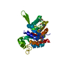
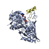
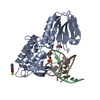
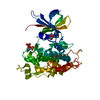
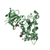
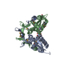
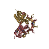
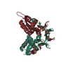
A: Putative transcription protein B: Putative transcription protein C: Putative transcription protein D: Putative transcription protein E: Putative transcription protein F: Putative transcription protein G: Putative transcription protein H: Putative transcription protein I: Putative transcription protein J: Putative transcription protein K: Putative transcription protein L: Putative transcription protein hetero molecules
Method to determine structure: SAD / Resolution: 2.3→29.72 Å / Cor.coef. Fo:Fc: 0.954 / Cor.coef. Fo:Fc free: 0.918 / SU B: 18.616 / SU ML: 0.211 / Cross valid method: THROUGHOUT / ESU R: 0.334 / ESU R Free: 0.269 / Stereochemistry target values: MAXIMUM LIKELIHOOD / Details: HYDROGENS HAVE BEEN ADDED IN THE RIDING POSITIONS
Rfactor
Num. reflection
% reflection
Selection details
Rfree
0.28695
4824
5 %
RANDOM
Rwork
0.21399
-
-
-
obs
0.21763
91451
99.06 %
-
Solvent computation
Ion probe radii: 0.8 Å / Shrinkage radii: 0.8 Å / VDW probe radii: 1.4 Å / Solvent model: MASK
Displacement parameters
Biso mean: 50.62 Å2
Baniso -1
Baniso -2
Baniso -3
1-
0.02 Å2
0 Å2
0 Å2
2-
-
0.01 Å2
0 Å2
3-
-
-
-0.03 Å2
Refinement step
Cycle: LAST / Resolution: 2.3→29.72 Å
Protein
Nucleic acid
Ligand
Solvent
Total
Num. atoms
13731
0
103
770
14604
Refine LS restraints
Refine-ID
Type
Dev ideal
Dev ideal target
Number
X-RAY DIFFRACTION
r_bond_refined_d
0.016
0.022
14068
X-RAY DIFFRACTION
r_bond_other_d
X-RAY DIFFRACTION
r_angle_refined_deg
1.771
1.963
18988
X-RAY DIFFRACTION
r_angle_other_deg
X-RAY DIFFRACTION
r_dihedral_angle_1_deg
6.141
5
1697
X-RAY DIFFRACTION
r_dihedral_angle_2_deg
27.881
21.798
712
X-RAY DIFFRACTION
r_dihedral_angle_3_deg
20.864
15
2402
X-RAY DIFFRACTION
r_dihedral_angle_4_deg
22.176
15
214
X-RAY DIFFRACTION
r_chiral_restr
0.133
0.2
2033
X-RAY DIFFRACTION
r_gen_planes_refined
0.008
0.021
10818
X-RAY DIFFRACTION
r_gen_planes_other
X-RAY DIFFRACTION
r_nbd_refined
X-RAY DIFFRACTION
r_nbd_other
X-RAY DIFFRACTION
r_nbtor_refined
X-RAY DIFFRACTION
r_nbtor_other
X-RAY DIFFRACTION
r_xyhbond_nbd_refined
X-RAY DIFFRACTION
r_xyhbond_nbd_other
X-RAY DIFFRACTION
r_metal_ion_refined
X-RAY DIFFRACTION
r_metal_ion_other
X-RAY DIFFRACTION
r_symmetry_vdw_refined
X-RAY DIFFRACTION
r_symmetry_vdw_other
X-RAY DIFFRACTION
r_symmetry_hbond_refined
X-RAY DIFFRACTION
r_symmetry_hbond_other
X-RAY DIFFRACTION
r_symmetry_metal_ion_refined
X-RAY DIFFRACTION
r_symmetry_metal_ion_other
X-RAY DIFFRACTION
r_mcbond_it
0.827
1.5
8546
X-RAY DIFFRACTION
r_mcbond_other
X-RAY DIFFRACTION
r_mcangle_it
1.618
2
13679
X-RAY DIFFRACTION
r_scbond_it
3.174
3
5522
X-RAY DIFFRACTION
r_scangle_it
5.073
4.5
5309
X-RAY DIFFRACTION
r_rigid_bond_restr
X-RAY DIFFRACTION
r_sphericity_free
X-RAY DIFFRACTION
r_sphericity_bonded
LS refinement shell
Resolution: 2.301→2.361 Å / Total num. of bins used: 20
Rfactor
Num. reflection
% reflection
Rfree
0.359
376
-
Rwork
0.289
6403
-
obs
-
-
96.43 %
Refinement TLS params.
Method: refined / Refine-ID: X-RAY DIFFRACTION
ID
L11 (°2)
L12 (°2)
L13 (°2)
L22 (°2)
L23 (°2)
L33 (°2)
S11 (Å °)
S12 (Å °)
S13 (Å °)
S21 (Å °)
S22 (Å °)
S23 (Å °)
S31 (Å °)
S32 (Å °)
S33 (Å °)
T11 (Å2)
T12 (Å2)
T13 (Å2)
T22 (Å2)
T23 (Å2)
T33 (Å2)
Origin x (Å)
Origin y (Å)
Origin z (Å)
1
2.2307
1.2894
1.1035
2.9903
0.6824
2.031
0.1486
0.0963
-0.3234
0.2092
-0.0502
-0.0891
0.268
-0.1061
-0.0984
0.0859
0.0572
-0.0286
0.1731
-0.0196
0.1519
60.597
-6.9743
147.2759
2
2.6684
0.7496
0.1795
3.4907
-0.1755
1.7476
0.0884
0.1749
0.1264
0.1767
-0.0779
0.0801
0.0091
-0.0222
-0.0105
0.0303
0.0556
-0.0027
0.1776
-0.0381
0.0871
63.7076
7.7245
146.5683
3
3.7669
-0.1503
-1.5977
0.8946
0.1567
2.5195
0.0859
-0.3361
0.1809
-0.1123
-0.0591
-0.1509
-0.0585
0.2144
-0.0267
0.1128
-0.0256
0.0029
0.2765
-0.0437
0.1383
40.1697
19.8509
114.3977
4
2.6594
-0.3522
0.5194
1.7686
-0.2227
2.0443
0.0766
-0.3068
-0.1125
-0.1759
-0.0356
-0.0023
0.0243
-0.0542
-0.041
0.0893
-0.0138
-0.0097
0.2843
0.0254
0.1321
27.3177
16.1676
113.8629
5
4.3007
0.0046
-1.5282
1.3344
0.4647
1.9046
0.1457
-0.2679
0.2377
-0.0749
-0.0632
-0.2157
-0.0117
0.2815
-0.0826
0.2076
0.0056
0.0331
0.058
-0.0054
0.1441
40.1649
19.6195
224.4735
6
3.6541
-0.2186
0.0309
2.3322
-0.006
1.5182
0.1115
-0.1651
-0.1112
-0.1734
-0.0917
0.0625
0.043
0.0388
-0.0198
0.169
0.0364
0.0334
0.0207
0.0177
0.0958
26.5505
15.3757
223.4642
7
1.5605
-0.3695
-0.0859
2.4998
0.6359
2.061
-0.2185
-0.0238
0.0378
0.1611
0.2715
-0.0913
0.0666
0.0306
-0.053
0.224
0.0904
-0.002
0.1714
-0.006
0.1314
37.1865
54.1768
190.8521
8
1.3992
-1.3192
0.4088
3.1667
-1.3426
3.0018
-0.2041
-0.019
0.0172
0.0826
0.2377
0.2777
-0.1333
-0.2312
-0.0336
0.1897
0.0578
0.0317
0.1669
0.0257
0.1602
26.6461
63.802
190.4174
9
1.5053
1.2791
0.9575
2.7886
1.0693
2.6851
0.1753
0.0906
-0.1963
0.2696
-0.1384
-0.047
0.198
-0.0609
-0.0368
0.2419
-0.0518
-0.032
0.1423
0.0101
0.1499
60.7671
-6.3707
37.6398
10
2.0446
0.6576
-0.4909
2.3734
-0.2953
2.0973
0.174
0.1159
0.0444
0.2956
-0.1414
0.1029
-0.0512
0.0238
-0.0326
0.2339
-0.0882
0.0155
0.1455
-0.0044
0.1261
64.0697
6.4708
37.0774
11
2.6711
-0.5586
-0.0342
3.4146
0.0319
1.4642
-0.1913
-0.0055
0.0183
-0.0251
0.209
-0.1344
0.0188
0.012
-0.0177
0.0775
-0.0798
-0.0265
0.1064
0.0244
0.0864
38.3294
53.9386
300.3271
12
2.0352
-1.4601
0.3644
4.126
-1.657
2.1504
-0.1882
-0.1043
0.0648
0.0595
0.2668
0.2801
-0.2421
-0.1894
-0.0786
0.1063
-0.06
-0.0175
0.1636
0.0304
0.1212
27.9085
63.7155
301.4393
Refinement TLS group
ID
Refine-ID
Refine TLS-ID
Auth asym-ID
Auth seq-ID
1
X-RAY DIFFRACTION
1
A
11 - 154
2
X-RAY DIFFRACTION
2
B
11 - 154
3
X-RAY DIFFRACTION
3
C
11 - 154
4
X-RAY DIFFRACTION
4
D
11 - 154
5
X-RAY DIFFRACTION
5
E
11 - 154
6
X-RAY DIFFRACTION
6
F
11 - 154
7
X-RAY DIFFRACTION
7
G
11 - 154
8
X-RAY DIFFRACTION
8
H
11 - 154
9
X-RAY DIFFRACTION
9
I
11 - 154
10
X-RAY DIFFRACTION
10
J
11 - 154
11
X-RAY DIFFRACTION
11
K
11 - 154
12
X-RAY DIFFRACTION
12
L
11 - 154
+
About Yorodumi
-
News
-
Feb 9, 2022. New format data for meta-information of EMDB entries
New format data for meta-information of EMDB entries
Version 3 of the EMDB header file is now the official format.
The previous official version 1.9 will be removed from the archive.
In the structure databanks used in Yorodumi, some data are registered as the other names, "COVID-19 virus" and "2019-nCoV". Here are the details of the virus and the list of structure data.
Jan 31, 2019. EMDB accession codes are about to change! (news from PDBe EMDB page)
EMDB accession codes are about to change! (news from PDBe EMDB page)
The allocation of 4 digits for EMDB accession codes will soon come to an end. Whilst these codes will remain in use, new EMDB accession codes will include an additional digit and will expand incrementally as the available range of codes is exhausted. The current 4-digit format prefixed with “EMD-” (i.e. EMD-XXXX) will advance to a 5-digit format (i.e. EMD-XXXXX), and so on. It is currently estimated that the 4-digit codes will be depleted around Spring 2019, at which point the 5-digit format will come into force.
The EM Navigator/Yorodumi systems omit the EMD- prefix.
Related info.:Q: What is EMD? / ID/Accession-code notation in Yorodumi/EM Navigator
Yorodumi is a browser for structure data from EMDB, PDB, SASBDB, etc.
This page is also the successor to EM Navigator detail page, and also detail information page/front-end page for Omokage search.
The word "yorodu" (or yorozu) is an old Japanese word meaning "ten thousand". "mi" (miru) is to see.
Related info.:EMDB / PDB / SASBDB / Comparison of 3 databanks / Yorodumi Search / Aug 31, 2016. New EM Navigator & Yorodumi / Yorodumi Papers / Jmol/JSmol / Function and homology information / Changes in new EM Navigator and Yorodumi
 Movie
Movie Controller

 Yorodumi
 Open data
 Basic information
 Components
 Keywords
 Function and homology information
 Pseudomonas aeruginosa PAO1 (bacteria)
 X-RAY DIFFRACTION /
  Authors
 Citation
 Structure visualization
 Downloads & links
 Other downloads


 PDBj
PDBj Assembly


 Mass: 22.990 Da / Num. of mol.: 11 / Source method: obtained synthetically / Formula: Na
 Mass: 92.094 Da / Num. of mol.: 12 / Source method: obtained synthetically / Formula: C3H8O3
 Mass: 94.971 Da / Num. of mol.: 4 / Source method: obtained synthetically / Formula: PO4
 Mass: 18.015 Da / Num. of mol.: 770 / Source method: isolated from a natural source / Formula: H2O
 Sample preparation
 / Beamline: 4A / Wavelength: 0.97959 Å
 Processing