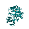
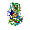
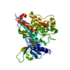
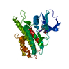
Glutathione conjugation / Estrogen biosynthesis / Synthesis of bile acids and bile salts via 24-hydroxycholesterol / Synthesis of bile acids and bile salts via 27-hydroxycholesterol / Synthesis of Prostaglandins (PG) and Thromboxanes (TX) / Formation of xylulose-5-phosphate / Prednisone ADME / Glycogen breakdown (glycogenolysis) / RA biosynthesis pathway / Retinoid metabolism and transport ...Glutathione conjugation / Estrogen biosynthesis / Synthesis of bile acids and bile salts via 24-hydroxycholesterol / Synthesis of bile acids and bile salts via 27-hydroxycholesterol / Synthesis of Prostaglandins (PG) and Thromboxanes (TX) / Formation of xylulose-5-phosphate / Prednisone ADME / Glycogen breakdown (glycogenolysis) / RA biosynthesis pathway / Retinoid metabolism and transport / Synthesis of bile acids and bile salts via 7alpha-hydroxycholesterol / aldose reductase (NADPH) activity / cytosol Similarity search - Function
Aldo/keto reductase family putative active site signature. / Aldo/keto reductase family signature 1. / NADP-dependent oxidoreductase domain / Aldo/keto reductase, conserved site / Aldo-keto reductase / NADP-dependent oxidoreductase domain / Aldo/keto reductase family / NADP-dependent oxidoreductase domain superfamily / TIM Barrel / Alpha-Beta Barrel / Alpha Beta Similarity search - Domain/homology
In the structure databanks used in Yorodumi, some data are registered as the other names, "COVID-19 virus" and "2019-nCoV". Here are the details of the virus and the list of structure data.
Jan 31, 2019. EMDB accession codes are about to change! (news from PDBe EMDB page)
EMDB accession codes are about to change! (news from PDBe EMDB page)
The allocation of 4 digits for EMDB accession codes will soon come to an end. Whilst these codes will remain in use, new EMDB accession codes will include an additional digit and will expand incrementally as the available range of codes is exhausted. The current 4-digit format prefixed with “EMD-” (i.e. EMD-XXXX) will advance to a 5-digit format (i.e. EMD-XXXXX), and so on. It is currently estimated that the 4-digit codes will be depleted around Spring 2019, at which point the 5-digit format will come into force.
The EM Navigator/Yorodumi systems omit the EMD- prefix.
Related info.:Q: What is EMD? / ID/Accession-code notation in Yorodumi/EM Navigator
Yorodumi is a browser for structure data from EMDB, PDB, SASBDB, etc.
This page is also the successor to EM Navigator detail page, and also detail information page/front-end page for Omokage search.
The word "yorodu" (or yorozu) is an old Japanese word meaning "ten thousand". "mi" (miru) is to see.
Related info.:EMDB / PDB / SASBDB / Comparison of 3 databanks / Yorodumi Search / Aug 31, 2016. New EM Navigator & Yorodumi / Yorodumi Papers / Jmol/JSmol / Function and homology information / Changes in new EM Navigator and Yorodumi
 Movie
Movie Controller

 Yorodumi
 Open data
 Basic information
 Components
 Keywords
 Function and homology information
 X-RAY DIFFRACTION /
  Authors
 Citation
 Structure visualization
 Downloads & links
 Other downloads
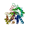


 PDBj
PDBj
 Assembly

 Mass: 18.015 Da / Num. of mol.: 330 / Source method: isolated from a natural source / Formula: H2O
 Sample preparation
 / Beamline: 22-ID / Wavelength: 1 Å
 Processing