Mass: 18.015 Da / Num. of mol.: 598 / Source method: isolated from a natural source / Formula: H2O
-
Details
Has protein modification
Y
Sequence details
THE CONSTRUCT WAS EXPRESSED WITH A PURIFICATION TAG MGSDKIHHHHHHENLYFQG. THE TAG WAS REMOVED WITH ...THE CONSTRUCT WAS EXPRESSED WITH A PURIFICATION TAG MGSDKIHHHHHHENLYFQG. THE TAG WAS REMOVED WITH TEV PROTEASE LEAVING ONLY A GLYCINE (0) FOLLOWED BY THE TARGET SEQUENCE.
-
Experimental details
-
Experiment
Experiment
Method: X-RAY DIFFRACTION / Number of used crystals: 1
-
Sample preparation
Crystal
Density Matthews: 2.85 Å3/Da / Density % sol: 56.79 %
Crystal grow
Temperature: 293 K / Method: vapor diffusion, sitting drop / pH: 7.5 Details: 10.00% Glycerol, 1.26M tri-Sodium Citrate, 0.1M HEPES pH 7.5, NANODROP, VAPOR DIFFUSION, SITTING DROP, temperature 293K
Monochromator: double crystal Si(111) / Protocol: SINGLE WAVELENGTH / Monochromatic (M) / Laue (L): M / Scattering type: x-ray
Radiation wavelength
Wavelength: 0.97915 Å / Relative weight: 1
Reflection
Resolution: 1.8→29.591 Å / Num. obs: 72528 / % possible obs: 98.2 % / Observed criterion σ(I): -3 / Biso Wilson estimate: 25.664 Å2 / Rmerge(I) obs: 0.062 / Net I/σ(I): 9.75
Reflection shell
Resolution (Å)
Rmerge(I) obs
Mean I/σ(I) obs
Num. measured obs
Num. unique obs
Diffraction-ID
% possible all
1.8-1.86
0.772
1.4
32242
12888
1
98.8
1.86-1.94
0.56
2
38183
14918
1
99.5
1.94-2.03
0.348
3.1
36294
14122
1
99.4
2.03-2.13
0.241
4.3
33298
12960
1
99.5
2.13-2.27
0.165
5.9
37592
14551
1
99.2
2.27-2.44
0.128
7.7
34735
13416
1
98.9
2.44-2.69
0.091
10.2
36101
13933
1
98.3
2.69-3.07
0.061
14.4
34913
13380
1
97.7
3.07-3.87
0.037
22.2
35301
13493
1
95.8
3.87-29.591
0.029
27.5
34823
13304
1
94.6
-
Phasing
Phasing
Method: SAD
-
Processing
Software
Name
Version
Classification
NB
MolProbity
3beta29
modelbuilding
PDB_EXTRACT
3.1
dataextraction
SHELX
phasing
SHARP
phasing
XSCALE
March15, 2012
datascaling
BUSTER-TNT
2.10.0
refinement
XDS
datareduction
SHELXD
phasing
BUSTER
2.10.0
refinement
Refinement
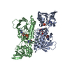
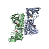
Method to determine structure: SAD / Resolution: 1.8→29.591 Å / Cor.coef. Fo:Fc: 0.9607 / Cor.coef. Fo:Fc free: 0.9556 / Occupancy max: 1 / Occupancy min: 0.23 / Cross valid method: THROUGHOUT / σ(F): 0 Details: 1. A MET-INHIBITION PROTOCOL WAS USED FOR SELENOMETHIONINE INCORPORATION DURING PROTEIN EXPRESSION. THE OCCUPANCY OF THE SE ATOMS IN THE MSE RESIDUES WAS REDUCED TO 0.75 FOR THE REDUCED ...Details: 1. A MET-INHIBITION PROTOCOL WAS USED FOR SELENOMETHIONINE INCORPORATION DURING PROTEIN EXPRESSION. THE OCCUPANCY OF THE SE ATOMS IN THE MSE RESIDUES WAS REDUCED TO 0.75 FOR THE REDUCED SCATTERING POWER DUE TO PARTIAL S-MET INCORPORATION. 2. ATOM RECORD CONTAINS SUM OF TLS AND RESIDUAL B FACTORS. ANISOU RECORD CONTAINS SUM OF TLS AND RESIDUAL U FACTORS. 3. THE MAD PHASES WERE USED AS RESTRAINTS DURING REFINEMENT. 4. HEPES, GLYCEROL, CHLORIDE MODELED ARE PRESENT IN CRYO CONDITIONS. FAD AND NADP ARE LIGANDS OF THE PROTEIN OBTAINED FROM THE EXPRESSION HOST. 5. NCS RESTRAINTS WERE APPLIED USING BUSTER'S LSSR RESTRAINT REPRESENTATION (-AUTONCS) 6. THE DISULFIDE BOND BETWEEN CYS134 AND CYS137 WAS MODELED AS PARTIALLY CLEAVED, LIKELY INDUCED BY RADIATION DAMAGE.
In the structure databanks used in Yorodumi, some data are registered as the other names, "COVID-19 virus" and "2019-nCoV". Here are the details of the virus and the list of structure data.
Jan 31, 2019. EMDB accession codes are about to change! (news from PDBe EMDB page)
EMDB accession codes are about to change! (news from PDBe EMDB page)
The allocation of 4 digits for EMDB accession codes will soon come to an end. Whilst these codes will remain in use, new EMDB accession codes will include an additional digit and will expand incrementally as the available range of codes is exhausted. The current 4-digit format prefixed with “EMD-” (i.e. EMD-XXXX) will advance to a 5-digit format (i.e. EMD-XXXXX), and so on. It is currently estimated that the 4-digit codes will be depleted around Spring 2019, at which point the 5-digit format will come into force.
The EM Navigator/Yorodumi systems omit the EMD- prefix.
Related info.:Q: What is EMD? / ID/Accession-code notation in Yorodumi/EM Navigator
Yorodumi is a browser for structure data from EMDB, PDB, SASBDB, etc.
This page is also the successor to EM Navigator detail page, and also detail information page/front-end page for Omokage search.
The word "yorodu" (or yorozu) is an old Japanese word meaning "ten thousand". "mi" (miru) is to see.
Related info.:EMDB / PDB / SASBDB / Comparison of 3 databanks / Yorodumi Search / Aug 31, 2016. New EM Navigator & Yorodumi / Yorodumi Papers / Jmol/JSmol / Function and homology information / Changes in new EM Navigator and Yorodumi
 Movie
Movie Controller

 Yorodumi
 Open data
 Basic information
 Components
 Keywords
 Function and homology information
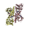
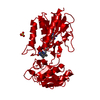
 Staphylococcus aureus subsp. aureus (bacteria)
 X-RAY DIFFRACTION /
  Authors
 Citation
 Structure visualization
 Downloads & links
 Other downloads
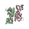

 PDBj
PDBj


 Assembly
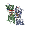


 Mass: 785.550 Da / Num. of mol.: 2 / Source method: obtained synthetically / Formula: C27H33N9O15P2 / Comment: FAD*YM
 Mass: 92.094 Da / Num. of mol.: 3 / Source method: obtained synthetically / Formula: C3H8O3
 Mass: 35.453 Da / Num. of mol.: 2 / Source method: obtained synthetically / Formula: Cl
 Mass: 743.405 Da / Num. of mol.: 1 / Source method: obtained synthetically / Formula: C21H28N7O17P3
 Mass: 238.305 Da / Num. of mol.: 1 / Source method: obtained synthetically / Formula: C8H18N2O4S / Comment: pH buffer*YM
 Sample preparation
 / Beamline: BL14-1 / Wavelength: 0.97915
  Processing