 Movie
Movie Controller

[English] 日本語
 Yorodumi
- PDB-4g6v: CdiA-CT/CdiI toxin and immunity complex from Burkholderia pseudomallei -
+ Open data

- Basic information
| Entry | Database: PDB / ID: 4g6v | ||||||
|---|---|---|---|---|---|---|---|
| Title | CdiA-CT/CdiI toxin and immunity complex from Burkholderia pseudomallei | ||||||
 Components Components |
| ||||||
 Keywords Keywords | TOXIN / tRNase / immunity | ||||||
| Function / homology |  Function and homology information Function and homology informationAlpha-Beta Plaits - #2920 / Trna Endonuclease; Chain: A, domain 1 - #120 / CDI inhibitor, Bp1026b-like / : / Trna Endonuclease; Chain: A, domain 1 / Alpha-Beta Plaits / 2-Layer Sandwich / 3-Layer(aba) Sandwich / Alpha Beta Similarity search - Domain/homology | ||||||
| Biological species |  Burkholderia pseudomallei 1026a (bacteria) Burkholderia pseudomallei 1026a (bacteria) | ||||||
| Method |  X-RAY DIFFRACTION / SYNCHROTRON / SAD / Resolution: 2.64 Å X-RAY DIFFRACTION / SYNCHROTRON / SAD / Resolution: 2.64 Å | ||||||
 Authors Authors | Morse, R.P. / Nikolakakis, K. / Willet, J. / Gerrick, E. / Low, D.A. / Hayes, C.S. / Goulding, C.W. | ||||||
 Citation Citation | Journal: Proc.Natl.Acad.Sci.USA / Year: 2012 Title: Structural basis of toxicity and immunity in contact-dependent growth inhibition (CDI) systems. Authors: Morse, R.P. / Nikolakakis, K.C. / Willett, J.L. / Gerrick, E. / Low, D.A. / Hayes, C.S. / Goulding, C.W. | ||||||
| History |
|
- Structure visualization
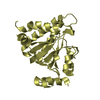
| Structure viewer | Molecule: MolmilJmol/JSmol |
|---|
- Downloads & links
-Download
| PDBx/mmCIF format | 4g6v.cif.gz | 182.4 KB | Display | PDBx/mmCIF format |
|---|---|---|---|---|
| PDB format | pdb4g6v.ent.gz | 145 KB | Display | PDB format |
| PDBx/mmJSON format | 4g6v.json.gz | Tree view | PDBx/mmJSON format | |
| Others |  Other downloads Other downloads |
-Validation report
| Summary document | 4g6v_validation.pdf.gz | 475.8 KB | Display | wwPDB validaton report |
|---|---|---|---|---|
| Full document | 4g6v_full_validation.pdf.gz | 486.7 KB | Display | |
| Data in XML | 4g6v_validation.xml.gz | 33.5 KB | Display | |
| Data in CIF | 4g6v_validation.cif.gz | 45.4 KB | Display | |
| Arichive directory | https://data.pdbj.org/pub/pdb/validation_reports/g6/4g6vftp://data.pdbj.org/pub/pdb/validation_reports/g6/4g6v | HTTPS FTP |
-Related structure data


-Links
 PDBj
PDBj

- Assembly
| Deposited unit | 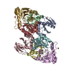
| |||||||||||||||||||||||||||||||||||||||||||||||||||||||||||
|---|---|---|---|---|---|---|---|---|---|---|---|---|---|---|---|---|---|---|---|---|---|---|---|---|---|---|---|---|---|---|---|---|---|---|---|---|---|---|---|---|---|---|---|---|---|---|---|---|---|---|---|---|---|---|---|---|---|---|---|---|
| 1 | 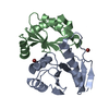
| |||||||||||||||||||||||||||||||||||||||||||||||||||||||||||
| 2 | 
| |||||||||||||||||||||||||||||||||||||||||||||||||||||||||||
| 3 | 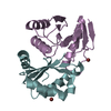
| |||||||||||||||||||||||||||||||||||||||||||||||||||||||||||
| 4 | 
| |||||||||||||||||||||||||||||||||||||||||||||||||||||||||||
| Unit cell |
| |||||||||||||||||||||||||||||||||||||||||||||||||||||||||||
| Noncrystallographic symmetry (NCS) | NCS domain:
NCS domain segments:
NCS ensembles :
|
-Components
| #1: Protein | Mass: 18228.092 Da / Num. of mol.: 4 / Fragment: UNP residues 2948-3122 Source method: isolated from a genetically manipulated source Source: (gene. exp.) Burkholderia pseudomallei 1026a (bacteria)Strain: 1026b / Gene: BP1026A_3896 / Production host: #2: Protein | Mass: 12474.045 Da / Num. of mol.: 4 Source method: isolated from a genetically manipulated source Source: (gene. exp.) Burkholderia pseudomallei 1026a (bacteria)Strain: 1026b / Gene: cdiI / Production host: #3: Chemical | ChemComp-BR /   Mass: 79.904 Da / Num. of mol.: 8 / Source method: obtained synthetically / Formula: Br Mass: 79.904 Da / Num. of mol.: 8 / Source method: obtained synthetically / Formula: Br#4: Water | ChemComp-HOH / |  Mass: 18.015 Da / Num. of mol.: 33 / Source method: isolated from a natural source / Formula: H2O Mass: 18.015 Da / Num. of mol.: 33 / Source method: isolated from a natural source / Formula: H2O |
|---|
-Experimental details
-Experiment
| Experiment | Method: X-RAY DIFFRACTION / Number of used crystals: 1 |
|---|
- Sample preparation
| Crystal | Density Matthews: 2.35 Å3/Da / Density % sol: 47.61 % |
|---|---|
| Crystal grow | Temperature: 298 K / Method: vapor diffusion, hanging drop / pH: 7.2 Details: 0.49 M sodium phosphate monobasic, 0.96 M potassium phosphate dibasic, 10 mM triethylene glycol, 504 nM chymotrypsin, pH 7.2, VAPOR DIFFUSION, HANGING DROP, temperature 298K |
-Data collection
| Diffraction | Mean temperature: 100 K |
|---|---|
| Diffraction source | Source: SYNCHROTRON / Site: ALS  / Beamline: 8.2.1 / Wavelength: 0.92 Å / Beamline: 8.2.1 / Wavelength: 0.92 Å |
| Detector | Type: ADSC QUANTUM 315r / Detector: CCD / Date: Jun 11, 2011 |
| Radiation | Monochromator: Double crystal, Si(111) / Protocol: SINGLE WAVELENGTH / Monochromatic (M) / Laue (L): M / Scattering type: x-ray |
| Radiation wavelength | Wavelength: 0.92 Å / Relative weight: 1 |
| Reflection | Resolution: 2.64→43.4 Å / Num. obs: 34071 / % possible obs: 99.47 % / Observed criterion σ(I): -3 / Redundancy: 29.6 % |
| Reflection shell | Resolution: 2.64→2.74 Å / Redundancy: 30.1 % / % possible all: 100 |
- Processing
| Software |
| |||||||||||||||||||||||||||||||||||||||||||||||||||||||||||||||||||||||||||||||||||||||||||
|---|---|---|---|---|---|---|---|---|---|---|---|---|---|---|---|---|---|---|---|---|---|---|---|---|---|---|---|---|---|---|---|---|---|---|---|---|---|---|---|---|---|---|---|---|---|---|---|---|---|---|---|---|---|---|---|---|---|---|---|---|---|---|---|---|---|---|---|---|---|---|---|---|---|---|---|---|---|---|---|---|---|---|---|---|---|---|---|---|---|---|---|---|
| Refinement | Method to determine structure: SAD / Resolution: 2.64→43.4 Å / SU ML: 0.76 / σ(F): 1.35 / Phase error: 27.62 / Stereochemistry target values: ML
| |||||||||||||||||||||||||||||||||||||||||||||||||||||||||||||||||||||||||||||||||||||||||||
| Solvent computation | Shrinkage radii: 0.72 Å / VDW probe radii: 1 Å / Solvent model: FLAT BULK SOLVENT MODEL / Bsol: 14.767 Å2 / ksol: 0.306 e/Å3 | |||||||||||||||||||||||||||||||||||||||||||||||||||||||||||||||||||||||||||||||||||||||||||
| Displacement parameters |
| |||||||||||||||||||||||||||||||||||||||||||||||||||||||||||||||||||||||||||||||||||||||||||
| Refinement step | Cycle: LAST / Resolution: 2.64→43.4 Å
| |||||||||||||||||||||||||||||||||||||||||||||||||||||||||||||||||||||||||||||||||||||||||||
| Refine LS restraints |
| |||||||||||||||||||||||||||||||||||||||||||||||||||||||||||||||||||||||||||||||||||||||||||
| Refine LS restraints NCS |
| |||||||||||||||||||||||||||||||||||||||||||||||||||||||||||||||||||||||||||||||||||||||||||
| LS refinement shell |
|
