 Movie
Movie Controller

[English] 日本語
 Yorodumi
- PDB-4fz2: Crystal structure of the fourth type of archaeal tRNA splicing en... -
+ Open data

- Basic information
| Entry | Database: PDB / ID: 4fz2 | ||||||
|---|---|---|---|---|---|---|---|
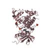
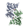
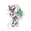
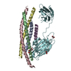
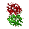
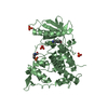
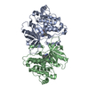
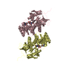
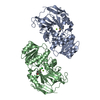
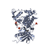
| Title | Crystal structure of the fourth type of archaeal tRNA splicing endonuclease from Candidatus Micrarchaeum acidiphilum ARMAN-2 | ||||||
 Components Components | tRNA intron endonuclease | ||||||
 Keywords Keywords | HYDROLASE / tRNA splicing endonuclease | ||||||
| Function / homology |  Function and homology information Function and homology informationtRNA-intron lyase activity / tRNA splicing, via endonucleolytic cleavage and ligation / nucleic acid binding / cytoplasm Similarity search - Function | ||||||
| Biological species |  Candidatus Micrarchaeum acidiphilum (archaea) Candidatus Micrarchaeum acidiphilum (archaea) | ||||||
| Method |  X-RAY DIFFRACTION / SYNCHROTRON / SAD / Resolution: 2.252 Å X-RAY DIFFRACTION / SYNCHROTRON / SAD / Resolution: 2.252 Å | ||||||
 Authors Authors | Hirata, A. / Fujishima, K. / Yamagami, R. / Kawamura, T. / Banfiled, J.F. / Kanai, A. / Hori, H. | ||||||
 Citation Citation | Journal: Nucleic Acids Res. / Year: 2012 Title: X-ray structure of the fourth type of archaeal tRNA splicing endonuclease: insights into the evolution of a novel three-unit composition and a unique loop involved in broad substrate specificity Authors: Hirata, A. / Fujishima, K. / Yamagami, R. / Kawamura, T. / Banfield, J.F. / Kanai, A. / Hori, H. | ||||||
| History |
|
- Structure visualization
| Structure viewer | Molecule: MolmilJmol/JSmol |
|---|
- Downloads & links
-Download
| PDBx/mmCIF format | 4fz2.cif.gz | 166.8 KB | Display | PDBx/mmCIF format |
|---|---|---|---|---|
| PDB format | pdb4fz2.ent.gz | 133.3 KB | Display | PDB format |
| PDBx/mmJSON format | 4fz2.json.gz | Tree view | PDBx/mmJSON format | |
| Others |  Other downloads Other downloads |
-Validation report
| Arichive directory | https://data.pdbj.org/pub/pdb/validation_reports/fz/4fz2ftp://data.pdbj.org/pub/pdb/validation_reports/fz/4fz2 | HTTPS FTP |
|---|
-Related structure data
| Similar structure data |
|---|
-Links
 PDBj
PDBj- Assembly
| Deposited unit | 
| ||||||||
|---|---|---|---|---|---|---|---|---|---|
| 1 |
| ||||||||
| Unit cell |
|
-Components
| #1: Protein | Mass: 45882.676 Da / Num. of mol.: 2 Source method: isolated from a genetically manipulated source Source: (gene. exp.) Candidatus Micrarchaeum acidiphilum (archaea)Strain: ARMAN-2 / Gene: UNLARM2_0797 / Plasmid: pET23b / Production host:  #2: Water | ChemComp-HOH / |  Mass: 18.015 Da / Num. of mol.: 152 / Source method: isolated from a natural source / Formula: H2O Mass: 18.015 Da / Num. of mol.: 152 / Source method: isolated from a natural source / Formula: H2O |
|---|
-Experimental details
-Experiment
| Experiment | Method: X-RAY DIFFRACTION / Number of used crystals: 1 |
|---|
- Sample preparation
| Crystal | Density Matthews: 3.2 Å3/Da / Density % sol: 61.57 % |
|---|---|
| Crystal grow | Temperature: 293 K / Method: vapor diffusion, hanging drop / pH: 7 Details: 18% PEG 3350, 0.2M tri-ammonium citrate, pH 7.0, VAPOR DIFFUSION, HANGING DROP, temperature 293K |
-Data collection
| Diffraction | Mean temperature: 100 K |
|---|---|
| Diffraction source | Source: SYNCHROTRON / Site: SPring-8  / Beamline: BL38B1 / Wavelength: 1 Å / Beamline: BL38B1 / Wavelength: 1 Å |
| Detector | Type: ADSC QUANTUM 315 / Detector: CCD / Date: Jun 25, 2012 |
| Radiation | Protocol: SINGLE WAVELENGTH / Monochromatic (M) / Laue (L): M / Scattering type: x-ray |
| Radiation wavelength | Wavelength: 1 Å / Relative weight: 1 |
| Reflection | Resolution: 2.25→50 Å / Num. obs: 53752 / % possible obs: 99.4 % / Observed criterion σ(F): 0 / Observed criterion σ(I): 0 / Redundancy: 5.9 % / Biso Wilson estimate: 39.01 Å2 / Rmerge(I) obs: 0.065 / Net I/σ(I): 39.2 |
| Reflection shell | Resolution: 2.25→2.33 Å / Redundancy: 5.9 % / Rmerge(I) obs: 0.065 / Mean I/σ(I) obs: 49.2 / % possible all: 99.4 |
- Processing
| Software |
| |||||||||||||||||||||||||||||||||||||||||||||||||||||||||||||||||||||||||||||||||||||||||||||||||||||||||||||||||||||||||||||||||||||
|---|---|---|---|---|---|---|---|---|---|---|---|---|---|---|---|---|---|---|---|---|---|---|---|---|---|---|---|---|---|---|---|---|---|---|---|---|---|---|---|---|---|---|---|---|---|---|---|---|---|---|---|---|---|---|---|---|---|---|---|---|---|---|---|---|---|---|---|---|---|---|---|---|---|---|---|---|---|---|---|---|---|---|---|---|---|---|---|---|---|---|---|---|---|---|---|---|---|---|---|---|---|---|---|---|---|---|---|---|---|---|---|---|---|---|---|---|---|---|---|---|---|---|---|---|---|---|---|---|---|---|---|---|---|---|
| Refinement | Method to determine structure: SAD / Resolution: 2.252→37.405 Å / Occupancy max: 1 / Occupancy min: 1 / FOM work R set: 0.7794 / SU ML: 1.79 / σ(F): 0.1 / Phase error: 29.29 / Stereochemistry target values: ML
| |||||||||||||||||||||||||||||||||||||||||||||||||||||||||||||||||||||||||||||||||||||||||||||||||||||||||||||||||||||||||||||||||||||
| Solvent computation | Shrinkage radii: 0.9 Å / VDW probe radii: 1.11 Å / Solvent model: FLAT BULK SOLVENT MODEL / Bsol: 51.545 Å2 / ksol: 0.367 e/Å3 | |||||||||||||||||||||||||||||||||||||||||||||||||||||||||||||||||||||||||||||||||||||||||||||||||||||||||||||||||||||||||||||||||||||
| Displacement parameters | Biso max: 129.61 Å2 / Biso mean: 48.8894 Å2 / Biso min: 23.57 Å2
| |||||||||||||||||||||||||||||||||||||||||||||||||||||||||||||||||||||||||||||||||||||||||||||||||||||||||||||||||||||||||||||||||||||
| Refinement step | Cycle: LAST / Resolution: 2.252→37.405 Å
| |||||||||||||||||||||||||||||||||||||||||||||||||||||||||||||||||||||||||||||||||||||||||||||||||||||||||||||||||||||||||||||||||||||
| Refine LS restraints |
| |||||||||||||||||||||||||||||||||||||||||||||||||||||||||||||||||||||||||||||||||||||||||||||||||||||||||||||||||||||||||||||||||||||
| LS refinement shell | Refine-ID: X-RAY DIFFRACTION / Total num. of bins used: 18
|
