- PDB-4fni: Crystal structure of IsdI-W66Y in complex with heme and cyanide -
+
Open data
ID or keywords:
Loading...
-
Basic information
Entry
Database: PDB / ID: 4fni
Title
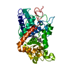
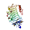
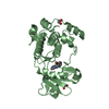
Crystal structure of IsdI-W66Y in complex with heme and cyanide
Components
Heme-degrading monooxygenase isdI
Keywords
OXIDOREDUCTASE / Dimeric alpha+beta barrel
Function / homology
Function and homology information
heme oxygenase (staphylobilin-producing) / iron import into cell / heme oxygenase (decyclizing) activity / heme catabolic process / iron ion binding / heme binding / cytoplasm Similarity search - Function
Mass: 18.015 Da / Num. of mol.: 338 / Source method: isolated from a natural source / Formula: H2O
-
Experimental details
-
Experiment
Experiment
Method: X-RAY DIFFRACTION / Number of used crystals: 1
-
Sample preparation
Crystal
Density Matthews: 2.66 Å3/Da / Density % sol: 53.76 %
Crystal grow
Temperature: 295 K / Method: vapor diffusion, hanging drop / pH: 5.5 Details: Crystals were grown in a reservoir solution containing 25% w/v polyethylene glycol 3350, 0.2 M magnesium chloride, and 0.1 M Bis-Tris, pH 5.5, and flash frozen with a cryoprotectant of 20% ...Details: Crystals were grown in a reservoir solution containing 25% w/v polyethylene glycol 3350, 0.2 M magnesium chloride, and 0.1 M Bis-Tris, pH 5.5, and flash frozen with a cryoprotectant of 20% ethylene glycol and 20 mM sodium cyanide solution., VAPOR DIFFUSION, HANGING DROP, temperature 295K
Resolution: 1.8→25.267 Å / Cor.coef. Fo:Fc: 0.943 / Cor.coef. Fo:Fc free: 0.924 / SU B: 2.719 / SU ML: 0.087 / Cross valid method: THROUGHOUT / ESU R: 0.145 / ESU R Free: 0.137 / Stereochemistry target values: MAXIMUM LIKELIHOOD / Details: HYDROGENS HAVE BEEN ADDED IN THE RIDING POSITIONS
Rfactor
Num. reflection
% reflection
Selection details
Rfree
0.23842
1288
5.1 %
RANDOM
Rwork
0.19727
-
-
-
all
0.19937
24090
-
-
obs
0.19937
24090
96.4 %
-
Solvent computation
Ion probe radii: 0.8 Å / Shrinkage radii: 0.8 Å / VDW probe radii: 1.4 Å / Solvent model: MASK
Displacement parameters
Biso mean: 18.886 Å2
Baniso -1
Baniso -2
Baniso -3
1-
-0.08 Å2
0 Å2
0 Å2
2-
-
0.06 Å2
0 Å2
3-
-
-
0.02 Å2
Refinement step
Cycle: LAST / Resolution: 1.8→25.267 Å
Protein
Nucleic acid
Ligand
Solvent
Total
Num. atoms
1830
0
91
338
2259
Refine LS restraints
Refine-ID
Type
Dev ideal
Dev ideal target
Number
X-RAY DIFFRACTION
r_bond_refined_d
0.012
0.023
2041
X-RAY DIFFRACTION
r_angle_refined_deg
1.187
1.996
2778
X-RAY DIFFRACTION
r_dihedral_angle_1_deg
5.962
5
236
X-RAY DIFFRACTION
r_dihedral_angle_2_deg
36.905
25.424
118
X-RAY DIFFRACTION
r_dihedral_angle_3_deg
12.139
15
367
X-RAY DIFFRACTION
r_dihedral_angle_4_deg
11.165
15
11
X-RAY DIFFRACTION
r_chiral_restr
0.076
0.2
271
X-RAY DIFFRACTION
r_gen_planes_refined
0.004
0.02
1609
X-RAY DIFFRACTION
r_mcbond_it
0.667
1.5
1129
X-RAY DIFFRACTION
r_mcangle_it
1.258
2
1828
X-RAY DIFFRACTION
r_scbond_it
1.753
3
912
X-RAY DIFFRACTION
r_scangle_it
2.813
4.5
943
LS refinement shell
Resolution: 1.8→1.847 Å / Total num. of bins used: 20
Rfactor
Num. reflection
% reflection
Rfree
0.353
72
-
Rwork
0.243
1262
-
obs
-
1262
70.88 %
+
About Yorodumi
-
News
-
Feb 9, 2022. New format data for meta-information of EMDB entries
New format data for meta-information of EMDB entries
Version 3 of the EMDB header file is now the official format.
The previous official version 1.9 will be removed from the archive.
In the structure databanks used in Yorodumi, some data are registered as the other names, "COVID-19 virus" and "2019-nCoV". Here are the details of the virus and the list of structure data.
Jan 31, 2019. EMDB accession codes are about to change! (news from PDBe EMDB page)
EMDB accession codes are about to change! (news from PDBe EMDB page)
The allocation of 4 digits for EMDB accession codes will soon come to an end. Whilst these codes will remain in use, new EMDB accession codes will include an additional digit and will expand incrementally as the available range of codes is exhausted. The current 4-digit format prefixed with “EMD-” (i.e. EMD-XXXX) will advance to a 5-digit format (i.e. EMD-XXXXX), and so on. It is currently estimated that the 4-digit codes will be depleted around Spring 2019, at which point the 5-digit format will come into force.
The EM Navigator/Yorodumi systems omit the EMD- prefix.
Related info.:Q: What is EMD? / ID/Accession-code notation in Yorodumi/EM Navigator
Yorodumi is a browser for structure data from EMDB, PDB, SASBDB, etc.
This page is also the successor to EM Navigator detail page, and also detail information page/front-end page for Omokage search.
The word "yorodu" (or yorozu) is an old Japanese word meaning "ten thousand". "mi" (miru) is to see.
Related info.:EMDB / PDB / SASBDB / Comparison of 3 databanks / Yorodumi Search / Aug 31, 2016. New EM Navigator & Yorodumi / Yorodumi Papers / Jmol/JSmol / Function and homology information / Changes in new EM Navigator and Yorodumi
 Movie
Movie Controller


 Open data
 Basic information
 Components
 Keywords
 Function and homology information
 Staphylococcus aureus subsp. aureus (bacteria)
 X-RAY DIFFRACTION /
  Authors
 Citation
 Structure visualization
 Downloads & links
 Other downloads


 PDBj
PDBj


 Assembly

 Mass: 616.487 Da / Num. of mol.: 2 / Source method: obtained synthetically / Formula: C34H32FeN4O4
 Mass: 26.017 Da / Num. of mol.: 2 / Source method: obtained synthetically / Formula: CN
 Mass: 24.305 Da / Num. of mol.: 1 / Source method: obtained synthetically / Formula: Mg
 Mass: 18.015 Da / Num. of mol.: 338 / Source method: isolated from a natural source / Formula: H2O
 Sample preparation
 Processing