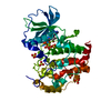
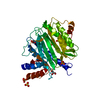
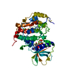
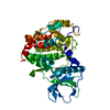
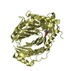
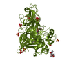
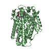
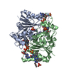
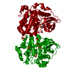
- PDB-4fi1: Crystal structure of scCK2 alpha in complex with ATP -
+
Open data
ID or keywords:
Loading...
-
Basic information
Entry
Database: PDB / ID: 4fi1
Title
Crystal structure of scCK2 alpha in complex with ATP
Components
Casein kinase II subunit alpha
Keywords
TRANSFERASE / protein-ATP complex / protein kinase / Phosphorylation
Function / homology
Function and homology information
donor selection / Receptor Mediated Mitophagy / Regulation of TP53 Activity through Phosphorylation / Condensation of Prometaphase Chromosomes / regulation of ribosomal protein gene transcription by RNA polymerase II / CURI complex / UTP-C complex / regulation of transcription by RNA polymerase III / regulation of transcription by RNA polymerase I / protein kinase CK2 complex ...donor selection / Receptor Mediated Mitophagy / Regulation of TP53 Activity through Phosphorylation / Condensation of Prometaphase Chromosomes / regulation of ribosomal protein gene transcription by RNA polymerase II / CURI complex / UTP-C complex / regulation of transcription by RNA polymerase III / regulation of transcription by RNA polymerase I / protein kinase CK2 complex / Regulation of PTEN stability and activity / nucleolar large rRNA transcription by RNA polymerase I / maturation of SSU-rRNA / small-subunit processome / ribosomal small subunit assembly / protein kinase activity / non-specific serine/threonine protein kinase / regulation of cell cycle / protein serine kinase activity / protein serine/threonine kinase activity / DNA damage response / nucleolus / nucleoplasm / ATP binding / nucleus / cytosol Similarity search - Function
Casein Kinase 2, subunit alpha / Phosphorylase Kinase; domain 1 / Phosphorylase Kinase; domain 1 / Transferase(Phosphotransferase) domain 1 / Transferase(Phosphotransferase); domain 1 / Serine/threonine-protein kinase, active site / Serine/Threonine protein kinases active-site signature. / Protein kinase domain / Serine/Threonine protein kinases, catalytic domain / Protein kinase, ATP binding site ...Casein Kinase 2, subunit alpha / Phosphorylase Kinase; domain 1 / Phosphorylase Kinase; domain 1 / Transferase(Phosphotransferase) domain 1 / Transferase(Phosphotransferase); domain 1 / Serine/threonine-protein kinase, active site / Serine/Threonine protein kinases active-site signature. / Protein kinase domain / Serine/Threonine protein kinases, catalytic domain / Protein kinase, ATP binding site / Protein kinases ATP-binding region signature. / Protein kinase domain profile. / Protein kinase domain / Protein kinase-like domain superfamily / 2-Layer Sandwich / Orthogonal Bundle / Mainly Alpha / Alpha Beta Similarity search - Domain/homology
Type: MAR scanner 345 mm plate / Detector: IMAGE PLATE / Date: Jan 1, 2011
Radiation
Scattering type: x-ray
Radiation wavelength
Wavelength: 0.9792 Å / Relative weight: 1
Reflection
Resolution: 2.09→60.43 Å / Num. obs: 29501
Reflection shell
Resolution: 2.095→2.149 Å / % possible all: 100
-
Processing
Software
Name
Version
Classification
HKL-2000
datacollection
REFMAC
5.6.0117
refinement
HKL-2000
datareduction
HKL-2000
datascaling
Refinement
Method to determine structure: MOLECULAR REPLACEMENT / Resolution: 2.09→60.43 Å / Cor.coef. Fo:Fc: 0.941 / Cor.coef. Fo:Fc free: 0.921 / SU B: 4.368 / SU ML: 0.118 / Cross valid method: THROUGHOUT / ESU R: 0.211 / ESU R Free: 0.181 / Stereochemistry target values: MAXIMUM LIKELIHOOD / Details: HYDROGENS HAVE BEEN USED IF PRESENT IN THE INPUT
Rfactor
Num. reflection
% reflection
Selection details
Rfree
0.24317
1452
5.1 %
RANDOM
Rwork
0.20432
-
-
-
obs
0.2063
27225
99.61 %
-
Solvent computation
Ion probe radii: 0.8 Å / Shrinkage radii: 0.8 Å / VDW probe radii: 1.2 Å / Solvent model: MASK
Displacement parameters
Biso mean: 25.464 Å2
Baniso -1
Baniso -2
Baniso -3
1-
0.58 Å2
0.29 Å2
-0 Å2
2-
-
0.58 Å2
-0 Å2
3-
-
-
-0.87 Å2
Refinement step
Cycle: LAST / Resolution: 2.09→60.43 Å
Protein
Nucleic acid
Ligand
Solvent
Total
Num. atoms
3070
0
63
141
3274
Refine LS restraints
Refine-ID
Type
Dev ideal
Dev ideal target
Number
X-RAY DIFFRACTION
r_bond_refined_d
0.007
0.02
3229
X-RAY DIFFRACTION
r_bond_other_d
X-RAY DIFFRACTION
r_angle_refined_deg
1.188
1.965
4382
X-RAY DIFFRACTION
r_angle_other_deg
X-RAY DIFFRACTION
r_dihedral_angle_1_deg
5.701
5
374
X-RAY DIFFRACTION
r_dihedral_angle_2_deg
35.183
23.846
156
X-RAY DIFFRACTION
r_dihedral_angle_3_deg
13.656
15
560
X-RAY DIFFRACTION
r_dihedral_angle_4_deg
15.732
15
18
X-RAY DIFFRACTION
r_chiral_restr
0.079
0.2
468
X-RAY DIFFRACTION
r_gen_planes_refined
0.005
0.021
2416
X-RAY DIFFRACTION
r_gen_planes_other
X-RAY DIFFRACTION
r_nbd_refined
X-RAY DIFFRACTION
r_nbd_other
X-RAY DIFFRACTION
r_nbtor_refined
X-RAY DIFFRACTION
r_nbtor_other
X-RAY DIFFRACTION
r_xyhbond_nbd_refined
X-RAY DIFFRACTION
r_xyhbond_nbd_other
X-RAY DIFFRACTION
r_metal_ion_refined
X-RAY DIFFRACTION
r_metal_ion_other
X-RAY DIFFRACTION
r_symmetry_vdw_refined
X-RAY DIFFRACTION
r_symmetry_vdw_other
X-RAY DIFFRACTION
r_symmetry_hbond_refined
X-RAY DIFFRACTION
r_symmetry_hbond_other
X-RAY DIFFRACTION
r_symmetry_metal_ion_refined
X-RAY DIFFRACTION
r_symmetry_metal_ion_other
X-RAY DIFFRACTION
r_mcbond_it
X-RAY DIFFRACTION
r_mcbond_other
X-RAY DIFFRACTION
r_mcangle_it
X-RAY DIFFRACTION
r_scbond_it
X-RAY DIFFRACTION
r_scangle_it
X-RAY DIFFRACTION
r_rigid_bond_restr
X-RAY DIFFRACTION
r_sphericity_free
X-RAY DIFFRACTION
r_sphericity_bonded
LS refinement shell
Resolution: 2.095→2.149 Å / Total num. of bins used: 20
Rfactor
Num. reflection
% reflection
Rfree
0.312
78
-
Rwork
0.22
1774
-
obs
-
-
96.91 %
+
About Yorodumi
-
News
-
Feb 9, 2022. New format data for meta-information of EMDB entries
New format data for meta-information of EMDB entries
Version 3 of the EMDB header file is now the official format.
The previous official version 1.9 will be removed from the archive.
In the structure databanks used in Yorodumi, some data are registered as the other names, "COVID-19 virus" and "2019-nCoV". Here are the details of the virus and the list of structure data.
Jan 31, 2019. EMDB accession codes are about to change! (news from PDBe EMDB page)
EMDB accession codes are about to change! (news from PDBe EMDB page)
The allocation of 4 digits for EMDB accession codes will soon come to an end. Whilst these codes will remain in use, new EMDB accession codes will include an additional digit and will expand incrementally as the available range of codes is exhausted. The current 4-digit format prefixed with “EMD-” (i.e. EMD-XXXX) will advance to a 5-digit format (i.e. EMD-XXXXX), and so on. It is currently estimated that the 4-digit codes will be depleted around Spring 2019, at which point the 5-digit format will come into force.
The EM Navigator/Yorodumi systems omit the EMD- prefix.
Related info.:Q: What is EMD? / ID/Accession-code notation in Yorodumi/EM Navigator
Yorodumi is a browser for structure data from EMDB, PDB, SASBDB, etc.
This page is also the successor to EM Navigator detail page, and also detail information page/front-end page for Omokage search.
The word "yorodu" (or yorozu) is an old Japanese word meaning "ten thousand". "mi" (miru) is to see.
Related info.:EMDB / PDB / SASBDB / Comparison of 3 databanks / Yorodumi Search / Aug 31, 2016. New EM Navigator & Yorodumi / Yorodumi Papers / Jmol/JSmol / Function and homology information / Changes in new EM Navigator and Yorodumi
 Movie
Movie Controller


 Open data
 Basic information
 Components
 Keywords
 Function and homology information
 X-RAY DIFFRACTION /
  Authors
 Citation
 Structure visualization
 Downloads & links
 Other downloads

 PDBj
PDBj


 Assembly
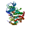


 Mass: 24.305 Da / Num. of mol.: 2 / Source method: obtained synthetically / Formula: Mg
 Mass: 507.181 Da / Num. of mol.: 1 / Source method: obtained synthetically / Formula: C10H16N5O13P3 / Comment: ATP, energy-carrying molecule*YM
 Mass: 96.063 Da / Num. of mol.: 6 / Source method: obtained synthetically / Formula: SO4
 Mass: 18.015 Da / Num. of mol.: 141 / Source method: isolated from a natural source / Formula: H2O
 Sample preparation
 Processing