 Movie
Movie Controller

[English] 日本語
 Yorodumi
- PDB-4fgc: Crystal Structure of Active Site Mutant C55A of Nitrile Reductase... -
+ Open data

- Basic information
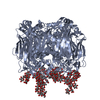
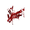
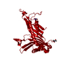
| Entry | Database: PDB / ID: 4fgc | ||||||
|---|---|---|---|---|---|---|---|
| Title | Crystal Structure of Active Site Mutant C55A of Nitrile Reductase QueF, Bound to Substrate PreQ0 | ||||||
 Components Components | NADPH-dependent 7-cyano-7-deazaguanine reductase | ||||||
 Keywords Keywords | OXIDOREDUCTASE / beta barrel / pterin binding fold / tunnel fold / tRNA modification enzyme / 7-cyano-7-deazaguanine (PreQ0) binding / NADPH binding | ||||||
| Function / homology |  Function and homology information Function and homology informationpreQ1 synthase / preQ1 synthase activity / tRNA modification / tRNA queuosine(34) biosynthetic process / metal ion binding / cytosol Similarity search - Function | ||||||
| Biological species |  | ||||||
| Method |  X-RAY DIFFRACTION / SYNCHROTRON / FOURIER SYNTHESIS / Resolution: 2.498 Å X-RAY DIFFRACTION / SYNCHROTRON / FOURIER SYNTHESIS / Resolution: 2.498 Å | ||||||
 Authors Authors | Stec, B. / Swairjo, M.A. | ||||||
 Citation Citation | Journal: J.Biol.Chem. / Year: 2012 Title: Structural basis of biological nitrile reduction. Authors: Chikwana, V.M. / Stec, B. / Lee, B.W. / de Crecy-Lagard, V. / Iwata-Reuyl, D. / Swairjo, M.A. | ||||||
| History |
|
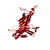
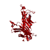
- Structure visualization
| Structure viewer | Molecule: MolmilJmol/JSmol |
|---|
- Downloads & links
-Download
| PDBx/mmCIF format | 4fgc.cif.gz | 167.4 KB | Display | PDBx/mmCIF format |
|---|---|---|---|---|
| PDB format | pdb4fgc.ent.gz | 132.3 KB | Display | PDB format |
| PDBx/mmJSON format | 4fgc.json.gz | Tree view | PDBx/mmJSON format | |
| Others |  Other downloads Other downloads |
-Validation report
| Arichive directory | https://data.pdbj.org/pub/pdb/validation_reports/fg/4fgcftp://data.pdbj.org/pub/pdb/validation_reports/fg/4fgc | HTTPS FTP |
|---|
-Related structure data
| Related structure data | 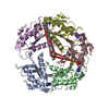 4f8bSC S: Starting model for refinement C: citing same article ( |
|---|---|
| Similar structure data |


-Links
 PDBj
PDBj
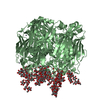
- Assembly
| Deposited unit | 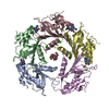
| ||||||||
|---|---|---|---|---|---|---|---|---|---|
| 1 | 
| ||||||||
| Unit cell |
|
-Components
| #1: Protein | Mass: 19364.891 Da / Num. of mol.: 5 / Mutation: C55A Source method: isolated from a genetically manipulated source Source: (gene. exp.) Strain: 168 / Gene: BSU13750, queF, ykvM / Plasmid: pET-30Xa / Production host: #2: Chemical | ChemComp-PQ0 / 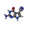  Mass: 175.147 Da / Num. of mol.: 4 / Source method: obtained synthetically / Formula: C7H5N5O Mass: 175.147 Da / Num. of mol.: 4 / Source method: obtained synthetically / Formula: C7H5N5O#3: Chemical | ChemComp-CA /   Mass: 40.078 Da / Num. of mol.: 8 / Source method: obtained synthetically / Formula: Ca Mass: 40.078 Da / Num. of mol.: 8 / Source method: obtained synthetically / Formula: Ca#4: Chemical | ChemComp-PE4 / |   Mass: 354.436 Da / Num. of mol.: 1 / Source method: obtained synthetically / Formula: C16H34O8 / Comment: precipitant*YM Mass: 354.436 Da / Num. of mol.: 1 / Source method: obtained synthetically / Formula: C16H34O8 / Comment: precipitant*YM#5: Water | ChemComp-HOH / |  Mass: 18.015 Da / Num. of mol.: 205 / Source method: isolated from a natural source / Formula: H2O Mass: 18.015 Da / Num. of mol.: 205 / Source method: isolated from a natural source / Formula: H2O |
|---|
-Experimental details
-Experiment
| Experiment | Method: X-RAY DIFFRACTION / Number of used crystals: 1 |
|---|
- Sample preparation
| Crystal | Density Matthews: 2.5 Å3/Da / Density % sol: 50.79 % |
|---|---|
| Crystal grow | Temperature: 293.15 K / Method: vapor diffusion / pH: 7.4 Details: 16-20% PEG500 MME, 60 mM imidazole, 40 mM imidazolium chloride, pH 7.4, 30 mM calcium chloride, VAPOR DIFFUSION, temperature 293.15K |
-Data collection
| Diffraction | Mean temperature: 200 K |
|---|---|
| Diffraction source | Source: SYNCHROTRON / Site: SSRL  / Beamline: BL9-1 / Wavelength: 1.000003 Å / Beamline: BL9-1 / Wavelength: 1.000003 Å |
| Detector | Type: ADSC QUANTUM 315r / Detector: CCD / Date: Feb 11, 2011 / Details: mirrors |
| Radiation | Monochromator: Side-scattering cuberoot I-beam bent single crystal, asymmetric cut 12.2 degrees Protocol: SINGLE WAVELENGTH / Monochromatic (M) / Laue (L): M / Scattering type: x-ray |
| Radiation wavelength | Wavelength: 1.000003 Å / Relative weight: 1 |
| Reflection | Resolution: 2.498→22.151 Å / Num. all: 34498 / Num. obs: 31577 / % possible obs: 91.6 % / Observed criterion σ(I): 0 / Redundancy: 4 % / Biso Wilson estimate: 46.6 Å2 / Rmerge(I) obs: 0.14 / Net I/σ(I): 6.8 |
| Reflection shell | Resolution: 2.498→2.54 Å / Redundancy: 3.9 % / Rmerge(I) obs: 0.65 / Mean I/σ(I) obs: 2.3 / Num. unique all: 1498 / % possible all: 88.8 |
- Processing
| Software |
| |||||||||||||||||||||||||||||||||||||||||||||||||||||||||||||||||
|---|---|---|---|---|---|---|---|---|---|---|---|---|---|---|---|---|---|---|---|---|---|---|---|---|---|---|---|---|---|---|---|---|---|---|---|---|---|---|---|---|---|---|---|---|---|---|---|---|---|---|---|---|---|---|---|---|---|---|---|---|---|---|---|---|---|---|
| Refinement | Method to determine structure: FOURIER SYNTHESIS Starting model: PDB ENTRY 4F8B Resolution: 2.498→22.15 Å / Cor.coef. Fo:Fc: 0.918 / Cor.coef. Fo:Fc free: 0.86 / SU B: 14.033 / SU ML: 0.304 / Isotropic thermal model: ISOTROPIC / Cross valid method: THROUGHOUT / ESU R: 0.711 / ESU R Free: 0.368 / Stereochemistry target values: MAXIMUM LIKELIHOOD Details: HYDROGENS HAVE BEEN ADDED IN THE RIDING POSITIONS U VALUES : REFINED INDIVIDUALLY
| |||||||||||||||||||||||||||||||||||||||||||||||||||||||||||||||||
| Solvent computation | Ion probe radii: 0.8 Å / Shrinkage radii: 0.8 Å / VDW probe radii: 1.4 Å / Solvent model: MASK | |||||||||||||||||||||||||||||||||||||||||||||||||||||||||||||||||
| Displacement parameters | Biso mean: 39.309 Å2
| |||||||||||||||||||||||||||||||||||||||||||||||||||||||||||||||||
| Refinement step | Cycle: LAST / Resolution: 2.498→22.15 Å
| |||||||||||||||||||||||||||||||||||||||||||||||||||||||||||||||||
| Refine LS restraints |
| |||||||||||||||||||||||||||||||||||||||||||||||||||||||||||||||||
| LS refinement shell | Resolution: 2.498→2.562 Å / Total num. of bins used: 20
|
