 Movie
Movie Controller

[English] 日本語
 Yorodumi
- PDB-4ffs: Crystal structure of 5'-methylthioadenosine/S-adenosylhomocystein... -
+ Open data

- Basic information
| Entry | Database: PDB / ID: 4ffs | ||||||
|---|---|---|---|---|---|---|---|
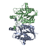
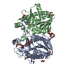
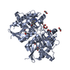
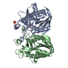
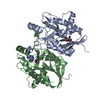
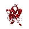
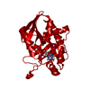
| Title | Crystal structure of 5'-methylthioadenosine/S-adenosylhomocysteine nucleosidase from Helicobacter pylori with butyl-thio-DADMe-Immucillin-A | ||||||
 Components Components | MTA/SAH nucleosidase | ||||||
 Keywords Keywords | HYDROLASE / L-methionine biosynthetic process from S-adenosylmethionine / L-methionine salvage from methylthioadenosine / 5'-methylthioadenosine/S-adenosylhomocysteine nucleosidase / adenosylhomocysteine nucleosidase activity / methylthioadenosine nucleosidase activity / hydrolase activity / acting on glycosyl bonds | ||||||
| Function / homology |  Function and homology information Function and homology informationaminodeoxyfutalosine nucleosidase / 6-amino-6-deoxyfutalosine hydrolase activity / adenosylhomocysteine nucleosidase / adenosylhomocysteine nucleosidase activity / methylthioadenosine nucleosidase activity / L-methionine salvage from S-adenosylmethionine / nucleoside catabolic process / L-methionine salvage from methylthioadenosine / menaquinone biosynthetic process / cytosol Similarity search - Function | ||||||
| Biological species |   Helicobacter pylori (bacteria) Helicobacter pylori (bacteria) | ||||||
| Method |  X-RAY DIFFRACTION / SYNCHROTRON / MOLECULAR REPLACEMENT / Resolution: 1.9 Å X-RAY DIFFRACTION / SYNCHROTRON / MOLECULAR REPLACEMENT / Resolution: 1.9 Å | ||||||
 Authors Authors | Haapalainen, A.M. / Rinaldo-Matthis, A. / Brown, R.L. / Norris, G.E. / Almo, S.C. / Schramm, V.L. | ||||||
 Citation Citation | Journal: Biochemistry / Year: 2012 Title: A Picomolar Transition State Analogue Inhibitor of MTAN as a Specific Antibiotic for Helicobacter pylori. Authors: Wang, S. / Haapalainen, A.M. / Yan, F. / Du, Q. / Tyler, P.C. / Evans, G.B. / Rinaldo-Matthis, A. / Brown, R.L. / Norris, G.E. / Almo, S.C. / Schramm, V.L. | ||||||
| History |
|
- Structure visualization
| Structure viewer | Molecule: MolmilJmol/JSmol |
|---|
- Downloads & links
-Download
| PDBx/mmCIF format | 4ffs.cif.gz | 64.9 KB | Display | PDBx/mmCIF format |
|---|---|---|---|---|
| PDB format | pdb4ffs.ent.gz | 47.9 KB | Display | PDB format |
| PDBx/mmJSON format | 4ffs.json.gz | Tree view | PDBx/mmJSON format | |
| Others |  Other downloads Other downloads |
-Validation report
| Summary document | 4ffs_validation.pdf.gz | 694.5 KB | Display | wwPDB validaton report |
|---|---|---|---|---|
| Full document | 4ffs_full_validation.pdf.gz | 694.9 KB | Display | |
| Data in XML | 4ffs_validation.xml.gz | 13 KB | Display | |
| Data in CIF | 4ffs_validation.cif.gz | 19.1 KB | Display | |
| Arichive directory | https://data.pdbj.org/pub/pdb/validation_reports/ff/4ffsftp://data.pdbj.org/pub/pdb/validation_reports/ff/4ffs | HTTPS FTP |
-Related structure data
| Similar structure data |
|---|


-Links
 PDBj
PDBj- Assembly
| Deposited unit | 
| |||||||||
|---|---|---|---|---|---|---|---|---|---|---|
| 1 | 
| |||||||||
| Unit cell |
| |||||||||
| Components on special symmetry positions |
|
-Components
| #1: Protein | Mass: 26286.268 Da / Num. of mol.: 1 Source method: isolated from a genetically manipulated source Source: (gene. exp.) Helicobacter pylori (bacteria) / Strain: J99 / Gene: jhp_0082, mtn, mtnN, Pfs / Plasmid: pET32a / Production host: References: UniProt: Q9ZMY2, adenosylhomocysteine nucleosidase |
|---|---|
| #2: Chemical | ChemComp-BIG / (  Mass: 335.468 Da / Num. of mol.: 1 / Source method: obtained synthetically / Formula: C16H25N5OS Mass: 335.468 Da / Num. of mol.: 1 / Source method: obtained synthetically / Formula: C16H25N5OS |
| #3: Chemical | ChemComp-CL /   Mass: 35.453 Da / Num. of mol.: 1 / Source method: obtained synthetically / Formula: Cl Mass: 35.453 Da / Num. of mol.: 1 / Source method: obtained synthetically / Formula: Cl |
| #4: Water | ChemComp-HOH /  Mass: 18.015 Da / Num. of mol.: 229 / Source method: isolated from a natural source / Formula: H2O Mass: 18.015 Da / Num. of mol.: 229 / Source method: isolated from a natural source / Formula: H2O |
-Experimental details
-Experiment
| Experiment | Method: X-RAY DIFFRACTION / Number of used crystals: 1 |
|---|
- Sample preparation
| Crystal | Density Matthews: 3.11 Å3/Da / Density % sol: 60.45 % |
|---|---|
| Crystal grow | Temperature: 293 K / Method: vapor diffusion, sitting drop / pH: 8.5 Details: 0.1M tri-sodium citrate, 0.1M Tris, pH 8.5, VAPOR DIFFUSION, SITTING DROP, temperature 293K |
-Data collection
| Diffraction | Mean temperature: 100 K |
|---|---|
| Diffraction source | Source: SYNCHROTRON / Site: NSLS  / Beamline: X29A / Wavelength: 1.1001 Å / Beamline: X29A / Wavelength: 1.1001 Å |
| Detector | Type: ADSC QUANTUM 315 / Detector: CCD / Date: Feb 16, 2006 / Details: MIRRORS |
| Radiation | Monochromator: SI III / Protocol: SINGLE WAVELENGTH / Monochromatic (M) / Laue (L): M / Scattering type: x-ray |
| Radiation wavelength | Wavelength: 1.1001 Å / Relative weight: 1 |
| Reflection | Resolution: 1.9→50 Å / Num. all: 26569 / Num. obs: 26569 / % possible obs: 99.9 % / Observed criterion σ(F): 0 / Observed criterion σ(I): 0 / Redundancy: 7 % / Biso Wilson estimate: 22.8 Å2 / Rmerge(I) obs: 0.099 / Rsym value: 0.099 / Net I/σ(I): 23.2 |
| Reflection shell | Resolution: 1.9→1.93 Å / Redundancy: 7.1 % / Rmerge(I) obs: 0.615 / Mean I/σ(I) obs: 2.9 / Num. unique all: 1301 / Rsym value: 0.615 / % possible all: 100 |
- Processing
| Software |
| ||||||||||||||||||||||||||||||||||||||||||||||||||||||||||||
|---|---|---|---|---|---|---|---|---|---|---|---|---|---|---|---|---|---|---|---|---|---|---|---|---|---|---|---|---|---|---|---|---|---|---|---|---|---|---|---|---|---|---|---|---|---|---|---|---|---|---|---|---|---|---|---|---|---|---|---|---|---|
| Refinement | Method to determine structure: MOLECULAR REPLACEMENT / Resolution: 1.9→42.19 Å / Cor.coef. Fo:Fc: 0.966 / Cor.coef. Fo:Fc free: 0.954 / SU B: 2.281 / SU ML: 0.068 / Cross valid method: THROUGHOUT / σ(F): 0 / σ(I): 0 / ESU R: 0.109 / ESU R Free: 0.106 / Stereochemistry target values: MAXIMUM LIKELIHOOD / Details: HYDROGENS HAVE BEEN ADDED IN THE RIDING POSITIONS
| ||||||||||||||||||||||||||||||||||||||||||||||||||||||||||||
| Solvent computation | Ion probe radii: 0.8 Å / Shrinkage radii: 0.8 Å / VDW probe radii: 1.2 Å / Solvent model: BABINET MODEL WITH MASK | ||||||||||||||||||||||||||||||||||||||||||||||||||||||||||||
| Displacement parameters | Biso mean: 26.016 Å2
| ||||||||||||||||||||||||||||||||||||||||||||||||||||||||||||
| Refinement step | Cycle: LAST / Resolution: 1.9→42.19 Å
| ||||||||||||||||||||||||||||||||||||||||||||||||||||||||||||
| Refine LS restraints |
| ||||||||||||||||||||||||||||||||||||||||||||||||||||||||||||
| LS refinement shell | Resolution: 1.901→1.95 Å / Total num. of bins used: 20
|
