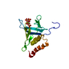
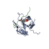
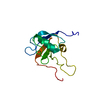
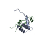
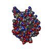
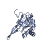
登録情報 データベース : PDB / ID : 4f7hタイトル The crystal structure of kindlin-2 pleckstrin homology domain in free form Fermitin family homolog 2 キーワード / / / / 機能・相同性 分子機能 ドメイン・相同性 構成要素
/ / / / / / / / / / / / / / / / / / / / / / / / / / / / / / / / / / / / / / / / / / / / / / / / / / / / / / / / / / / / / / / / / / / / / / / / / / / / / / / / / / / / / / / / / / 生物種 Homo sapiens (ヒト)手法 / / 解像度 : 1.9 Å データ登録者 Liu, Y. / Zhu, Y. / Qin, J. / Ye, S. / Zhang, R. ジャーナル : Protein Cell / 年 : 2012タイトル : Crystal structure of kindlin-2 PH domain reveals a conformational transition for its membrane anchoring and regulation of integrin activation.著者 : Liu, Y. / Zhu, Y. / Ye, S. / Zhang, R. 履歴 登録 2012年5月16日 登録サイト / 処理サイト 改定 1.0 2012年6月13日 Provider / タイプ 改定 1.1 2012年7月18日 Group 改定 1.2 2023年9月13日 Group Data collection / Database references ... Data collection / Database references / Derived calculations / Refinement description カテゴリ chem_comp_atom / chem_comp_bond ... chem_comp_atom / chem_comp_bond / database_2 / pdbx_initial_refinement_model / struct_ref_seq_dif / struct_site Item _database_2.pdbx_DOI / _database_2.pdbx_database_accession ... _database_2.pdbx_DOI / _database_2.pdbx_database_accession / _struct_ref_seq_dif.details / _struct_site.pdbx_auth_asym_id / _struct_site.pdbx_auth_comp_id / _struct_site.pdbx_auth_seq_id
すべて表示 表示を減らす
 ムービー
ムービー コントローラー


 データを開く
 基本情報
基本情報 要素
要素 キーワード
キーワード 機能・相同性情報
 Homo sapiens (ヒト)
 X線回折 /
  データ登録者
 引用
引用 構造の表示
構造の表示 ダウンロードとリンク
 その他のダウンロード


 PDBj
PDBj


 集合体
集合体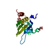


 分子量: 150.087 Da / 分子数: 2 / 由来タイプ: 合成 / 式: C4H6O6
 分子量: 18.015 Da / 分子数: 86 / 由来タイプ: 天然 / 式: H2O
 試料調製
試料調製 解析
解析