 Movie
Movie Controller

[English] 日本語
 Yorodumi
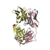
- PDB-4f7e: Crystal structure of bovine CD1d with bound C16:0-alpha-galactosy... -
+ Open data

- Basic information
| Entry | Database: PDB / ID: 4f7e | |||||||||
|---|---|---|---|---|---|---|---|---|---|---|
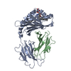
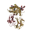
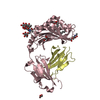
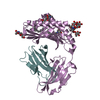
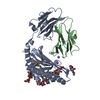
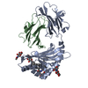
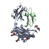
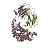
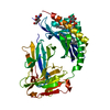
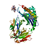
| Title | Crystal structure of bovine CD1d with bound C16:0-alpha-galactosyl ceramide | |||||||||
 Components Components |
| |||||||||
 Keywords Keywords | IMMUNE SYSTEM / protein-glycolipid complex / MHC-fold / Ig-fold / antigen presentation / TCR / membrane | |||||||||
| Function / homology |  Function and homology information Function and homology informationEndosomal/Vacuolar pathway / Antigen Presentation: Folding, assembly and peptide loading of class I MHC / Immunoregulatory interactions between a Lymphoid and a non-Lymphoid cell / ER-Phagosome pathway / DAP12 signaling / Neutrophil degranulation / response to metal ion / antigen processing and presentation of peptide antigen via MHC class I / MHC class I protein complex / peptide antigen assembly with MHC class II protein complex ...Endosomal/Vacuolar pathway / Antigen Presentation: Folding, assembly and peptide loading of class I MHC / Immunoregulatory interactions between a Lymphoid and a non-Lymphoid cell / ER-Phagosome pathway / DAP12 signaling / Neutrophil degranulation / response to metal ion / antigen processing and presentation of peptide antigen via MHC class I / MHC class I protein complex / peptide antigen assembly with MHC class II protein complex / MHC class II protein complex / antigen processing and presentation of exogenous peptide antigen via MHC class II / positive regulation of immune response / peptide antigen binding / positive regulation of T cell activation / MHC class II protein complex binding / late endosome membrane / endosome membrane / immune response / innate immune response / lysosomal membrane / extracellular region / plasma membrane Similarity search - Function | |||||||||
| Biological species |  | |||||||||
| Method |  X-RAY DIFFRACTION / SYNCHROTRON / MOLECULAR REPLACEMENT / molecular replacement / Resolution: 2.4 Å X-RAY DIFFRACTION / SYNCHROTRON / MOLECULAR REPLACEMENT / molecular replacement / Resolution: 2.4 Å | |||||||||
 Authors Authors | Wang, J. / Zajonc, D.M. | |||||||||
 Citation Citation | Journal: Plos One / Year: 2012 Title: Crystal Structures of Bovine CD1d Reveal Altered αGalCer Presentation and a Restricted A' Pocket Unable to Bind Long-Chain Glycolipids. Authors: Wang, J. / Guillaume, J. / Pauwels, N. / Van Calenbergh, S. / Van Rhijn, I. / Zajonc, D.M. | |||||||||
| History |
|
- Structure visualization
| Structure viewer | Molecule: MolmilJmol/JSmol |
|---|
- Downloads & links
-Download
| PDBx/mmCIF format | 4f7e.cif.gz | 93.4 KB | Display | PDBx/mmCIF format |
|---|---|---|---|---|
| PDB format | pdb4f7e.ent.gz | 68.6 KB | Display | PDB format |
| PDBx/mmJSON format | 4f7e.json.gz | Tree view | PDBx/mmJSON format | |
| Others |  Other downloads Other downloads |
-Validation report
| Arichive directory | https://data.pdbj.org/pub/pdb/validation_reports/f7/4f7eftp://data.pdbj.org/pub/pdb/validation_reports/f7/4f7e | HTTPS FTP |
|---|
-Related structure data
| Related structure data |  4f7cC  3l9rS C: citing same article ( S: Starting model for refinement |
|---|---|
| Similar structure data |


-Links
 PDBj
PDBj- Assembly
| Deposited unit | 
| ||||||||
|---|---|---|---|---|---|---|---|---|---|
| 1 |
| ||||||||
| Unit cell |
|
-Components
-Protein , 2 types, 2 molecules AB
| #1: Protein | Mass: 32460.561 Da / Num. of mol.: 1 / Fragment: UNP residues 129-405 Source method: isolated from a genetically manipulated source Source: (gene. exp.)   Spodoptera frugiperda (fall armyworm) / Strain (production host): SF9 / References: UniProt: A1L565 Spodoptera frugiperda (fall armyworm) / Strain (production host): SF9 / References: UniProt: A1L565 |
|---|---|
| #2: Protein | Mass: 11653.148 Da / Num. of mol.: 1 / Fragment: UNP residues 21-118 Source method: isolated from a genetically manipulated source Source: (gene. exp.) Spodoptera frugiperda (fall armyworm) / Strain (production host): SF9 / References: UniProt: P01888 |
-Sugars , 2 types, 2 molecules 
| #3: Polysaccharide | 2-acetamido-2-deoxy-beta-D-glucopyranose-(1-4)-2-acetamido-2-deoxy-beta-D-glucopyranose Source method: isolated from a genetically manipulated source |
|---|---|
| #5: Sugar | ChemComp-NAG /  Type: D-saccharide, beta linking / Mass: 221.208 Da / Num. of mol.: 1 Type: D-saccharide, beta linking / Mass: 221.208 Da / Num. of mol.: 1Source method: isolated from a genetically manipulated source Formula: C8H15NO6 |
-Non-polymers , 3 types, 28 molecules 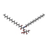


| #4: Chemical | ChemComp-0SH /  Mass: 718.057 Da / Num. of mol.: 1 / Source method: obtained synthetically / Formula: C40H79NO9 Mass: 718.057 Da / Num. of mol.: 1 / Source method: obtained synthetically / Formula: C40H79NO9 |
|---|---|
| #6: Chemical | ChemComp-SO4 /  Mass: 96.063 Da / Num. of mol.: 1 / Source method: obtained synthetically / Formula: SO4 Mass: 96.063 Da / Num. of mol.: 1 / Source method: obtained synthetically / Formula: SO4 |
| #7: Water | ChemComp-HOH / Mass: 18.015 Da / Num. of mol.: 26 / Source method: isolated from a natural source / Formula: H2O |
-Details
| Has protein modification | Y |
|---|
-Experimental details
-Experiment
| Experiment | Method: X-RAY DIFFRACTION / Number of used crystals: 1 |
|---|
- Sample preparation
| Crystal | Density Matthews: 2.9 Å3/Da / Density % sol: 57.54 % |
|---|---|
| Crystal grow | Temperature: 295 K / Method: vapor diffusion, sitting drop Details: 20% PEG4000, 200 mM potassium sulfate, VAPOR DIFFUSION, SITTING DROP, temperature 295K |
-Data collection
| Diffraction | Mean temperature: 100 K | ||||||||||||||||||||||||||||||||||||||||||||||||||||||||||||||||||||||||||||||||||||||||||||||||||||||||||||||||
|---|---|---|---|---|---|---|---|---|---|---|---|---|---|---|---|---|---|---|---|---|---|---|---|---|---|---|---|---|---|---|---|---|---|---|---|---|---|---|---|---|---|---|---|---|---|---|---|---|---|---|---|---|---|---|---|---|---|---|---|---|---|---|---|---|---|---|---|---|---|---|---|---|---|---|---|---|---|---|---|---|---|---|---|---|---|---|---|---|---|---|---|---|---|---|---|---|---|---|---|---|---|---|---|---|---|---|---|---|---|---|---|---|---|
| Diffraction source | Source: SYNCHROTRON / Site: SSRL  / Beamline: BL11-1 / Wavelength: 1 Å / Beamline: BL11-1 / Wavelength: 1 Å | ||||||||||||||||||||||||||||||||||||||||||||||||||||||||||||||||||||||||||||||||||||||||||||||||||||||||||||||||
| Detector | Type: DECTRIS PILATUS 6M / Detector: PIXEL / Date: Feb 3, 2012 / Details: Rh coated flat mirror | ||||||||||||||||||||||||||||||||||||||||||||||||||||||||||||||||||||||||||||||||||||||||||||||||||||||||||||||||
| Radiation | Monochromator: Side scattering bent cube-root I-beam single crystal, asymmetric cut 4.965 degrees Protocol: SINGLE WAVELENGTH / Monochromatic (M) / Laue (L): M / Scattering type: x-ray | ||||||||||||||||||||||||||||||||||||||||||||||||||||||||||||||||||||||||||||||||||||||||||||||||||||||||||||||||
| Radiation wavelength | Wavelength: 1 Å / Relative weight: 1 | ||||||||||||||||||||||||||||||||||||||||||||||||||||||||||||||||||||||||||||||||||||||||||||||||||||||||||||||||
| Reflection | Redundancy: 3.9 % / Number: 108859 / Rmerge(I) obs: 0.059 / Χ2: 1.57 / D res high: 2.15 Å / D res low: 40 Å / Num. obs: 28064 / % possible obs: 96.1 | ||||||||||||||||||||||||||||||||||||||||||||||||||||||||||||||||||||||||||||||||||||||||||||||||||||||||||||||||
| Diffraction reflection shell |
| ||||||||||||||||||||||||||||||||||||||||||||||||||||||||||||||||||||||||||||||||||||||||||||||||||||||||||||||||
| Reflection | Resolution: 2.4→40 Å / Num. all: 20240 / Num. obs: 20240 / % possible obs: 97.4 % / Observed criterion σ(F): 0 / Observed criterion σ(I): 0 / Redundancy: 4.1 % / Biso Wilson estimate: 45.6 Å2 / Rmerge(I) obs: 0.067 / Net I/σ(I): 35 | ||||||||||||||||||||||||||||||||||||||||||||||||||||||||||||||||||||||||||||||||||||||||||||||||||||||||||||||||
| Reflection shell | Resolution: 2.4→2.46 Å / Redundancy: 4.1 % / Rmerge(I) obs: 0.199 / Mean I/σ(I) obs: 4.9 / Num. unique all: 1901 / % possible all: 98.2 |
-Phasing
| Phasing | Method: molecular replacement |
|---|
- Processing
| Software |
| |||||||||||||||||||||||||||||||||||||||||||||
|---|---|---|---|---|---|---|---|---|---|---|---|---|---|---|---|---|---|---|---|---|---|---|---|---|---|---|---|---|---|---|---|---|---|---|---|---|---|---|---|---|---|---|---|---|---|---|
| Refinement | Method to determine structure: MOLECULAR REPLACEMENT Starting model: PDB ENTRY 3L9R Resolution: 2.4→35.9 Å / Cor.coef. Fo:Fc: 0.949 / Cor.coef. Fo:Fc free: 0.918 / WRfactor Rfree: 0.2878 / WRfactor Rwork: 0.2328 / Occupancy max: 1 / Occupancy min: 1 / FOM work R set: 0.7586 / SU B: 9.739 / SU ML: 0.218 / SU R Cruickshank DPI: 0.3582 / SU Rfree: 0.2704 / Cross valid method: THROUGHOUT / σ(F): 0 / ESU R: 0.358 / ESU R Free: 0.27 / Stereochemistry target values: MAXIMUM LIKELIHOOD Details: HYDROGENS HAVE BEEN USED IF PRESENT IN THE INPUT U VALUES : REFINED INDIVIDUALLY
| |||||||||||||||||||||||||||||||||||||||||||||
| Solvent computation | Ion probe radii: 0.8 Å / Shrinkage radii: 0.8 Å / VDW probe radii: 1.2 Å / Solvent model: MASK | |||||||||||||||||||||||||||||||||||||||||||||
| Displacement parameters | Biso max: 132.44 Å2 / Biso mean: 61.7012 Å2 / Biso min: 30.29 Å2
| |||||||||||||||||||||||||||||||||||||||||||||
| Refinement step | Cycle: LAST / Resolution: 2.4→35.9 Å
| |||||||||||||||||||||||||||||||||||||||||||||
| Refine LS restraints |
| |||||||||||||||||||||||||||||||||||||||||||||
| LS refinement shell | Resolution: 2.4→2.462 Å / Total num. of bins used: 20
|
