 Movie
Movie Controller


+ Open data

- Basic information
| Entry | Database: PDB / ID: 4f4l | ||||||
|---|---|---|---|---|---|---|---|
| Title | Open Channel Conformation of a Voltage Gated Sodium Channel | ||||||
 Components Components | Ion transport protein | ||||||
 Keywords Keywords | METAL TRANSPORT / alpha helical membrane protein / voltage-gated sodium channel / membrane | ||||||
| Function / homology |  Function and homology information Function and homology informationvoltage-gated sodium channel complex / voltage-gated sodium channel activity Similarity search - Function | ||||||
| Biological species |  Magnetococcus marinus (bacteria) Magnetococcus marinus (bacteria) | ||||||
| Method |  X-RAY DIFFRACTION / SYNCHROTRON / MOLECULAR REPLACEMENT / Resolution: 3.49 Å X-RAY DIFFRACTION / SYNCHROTRON / MOLECULAR REPLACEMENT / Resolution: 3.49 Å | ||||||
 Authors Authors | McCusker, E.C. / Bagneris, C. / Naylor, C.E. / Cole, A.R. / D'Avanzo, N. / Nichols, C.G. / Wallace, B.A. | ||||||
 Citation Citation | Journal: Nat Commun / Year: 2012 Title: Structure of a bacterial voltage-gated sodium channel pore reveals mechanisms of opening and closing. Authors: McCusker, E.C. / Bagneris, C. / Naylor, C.E. / Cole, A.R. / D'Avanzo, N. / Nichols, C.G. / Wallace, B.A. | ||||||
| History |
|
- Structure visualization
| Structure viewer | Molecule: MolmilJmol/JSmol |
|---|
- Downloads & links
-Download
| PDBx/mmCIF format | 4f4l.cif.gz | 80.7 KB | Display | PDBx/mmCIF format |
|---|---|---|---|---|
| PDB format | pdb4f4l.ent.gz | 60.9 KB | Display | PDB format |
| PDBx/mmJSON format | 4f4l.json.gz | Tree view | PDBx/mmJSON format | |
| Others |  Other downloads Other downloads |
-Validation report
| Summary document | 4f4l_validation.pdf.gz | 455.2 KB | Display | wwPDB validaton report |
|---|---|---|---|---|
| Full document | 4f4l_full_validation.pdf.gz | 462 KB | Display | |
| Data in XML | 4f4l_validation.xml.gz | 14.8 KB | Display | |
| Data in CIF | 4f4l_validation.cif.gz | 20.3 KB | Display | |
| Arichive directory | https://data.pdbj.org/pub/pdb/validation_reports/f4/4f4lftp://data.pdbj.org/pub/pdb/validation_reports/f4/4f4l | HTTPS FTP |
-Related structure data
| Related structure data | 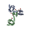 3rvyS S: Starting model for refinement |
|---|---|
| Similar structure data |


-Links
 PDBj
PDBj
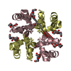
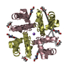
- Assembly
| Deposited unit | 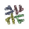
| ||||||||
|---|---|---|---|---|---|---|---|---|---|
| 1 |
| ||||||||
| Unit cell |
|
-Components
| #1: Protein | Mass: 12439.515 Da / Num. of mol.: 4 / Fragment: pore region (S5-C-terminal domain) Source method: isolated from a genetically manipulated source Source: (gene. exp.) Magnetococcus marinus (bacteria) / Strain: MC-1 / Gene: A0L5S6, Mmc1_0798 / Plasmid: pET15b / Production host: #2: Water | ChemComp-HOH / |  Mass: 18.015 Da / Num. of mol.: 42 / Source method: isolated from a natural source / Formula: H2O Mass: 18.015 Da / Num. of mol.: 42 / Source method: isolated from a natural source / Formula: H2O |
|---|
-Experimental details
-Experiment
| Experiment | Method: X-RAY DIFFRACTION / Number of used crystals: 2 |
|---|
- Sample preparation
| Crystal | Density Matthews: 4.14 Å3/Da / Density % sol: 70.31 % |
|---|---|
| Crystal grow | Temperature: 277 K / Method: vapor diffusion / pH: 5.6 Details: 30% PEG 400, 0.09 M tri-sodium citrate, pH 5.6, VAPOR DIFFUSION, temperature 277K |
-Data collection
| Diffraction | Mean temperature: 100 K | ||||||||||||||||||||||||||||||||||||||||||
|---|---|---|---|---|---|---|---|---|---|---|---|---|---|---|---|---|---|---|---|---|---|---|---|---|---|---|---|---|---|---|---|---|---|---|---|---|---|---|---|---|---|---|---|
| Diffraction source | Source: SYNCHROTRON / Site: ESRF  / Beamline: ID23-2 / Wavelength: 0.8726 Å / Beamline: ID23-2 / Wavelength: 0.8726 Å | ||||||||||||||||||||||||||||||||||||||||||
| Detector | Type: MARMOSAIC 225 mm CCD / Detector: CCD / Date: Nov 23, 2011 / Details: Si crystal | ||||||||||||||||||||||||||||||||||||||||||
| Radiation | Protocol: SINGLE WAVELENGTH / Monochromatic (M) / Laue (L): M / Scattering type: x-ray | ||||||||||||||||||||||||||||||||||||||||||
| Radiation wavelength | Wavelength: 0.8726 Å / Relative weight: 1 | ||||||||||||||||||||||||||||||||||||||||||
| Reflection | Resolution: 3.49→95.1 Å / Num. all: 10528 / Num. obs: 8747 / % possible obs: 83.1 % / Observed criterion σ(F): 0 / Observed criterion σ(I): 0 / Redundancy: 11.1 % / Biso Wilson estimate: 92.44 Å2 / Rmerge(I) obs: 0.048 / Rsym value: 0.05 / Net I/σ(I): 22.98 | ||||||||||||||||||||||||||||||||||||||||||
| Reflection shell | Diffraction-ID: 1
|
- Processing
| Software |
| ||||||||||||||||||||||||||||||||||||||||||||||||||||||||||||||||||
|---|---|---|---|---|---|---|---|---|---|---|---|---|---|---|---|---|---|---|---|---|---|---|---|---|---|---|---|---|---|---|---|---|---|---|---|---|---|---|---|---|---|---|---|---|---|---|---|---|---|---|---|---|---|---|---|---|---|---|---|---|---|---|---|---|---|---|---|
| Refinement | Method to determine structure: MOLECULAR REPLACEMENT Starting model: PDB ENTRY 3RVY Resolution: 3.49→43.62 Å / Cor.coef. Fo:Fc: 0.8956 / Cor.coef. Fo:Fc free: 0.8951 / Cross valid method: THROUGHOUT / σ(F): 0 / Stereochemistry target values: Engh & Huber
| ||||||||||||||||||||||||||||||||||||||||||||||||||||||||||||||||||
| Displacement parameters | Biso mean: 137.98 Å2
| ||||||||||||||||||||||||||||||||||||||||||||||||||||||||||||||||||
| Refine analyze |
| ||||||||||||||||||||||||||||||||||||||||||||||||||||||||||||||||||
| Refinement step | Cycle: LAST / Resolution: 3.49→43.62 Å
| ||||||||||||||||||||||||||||||||||||||||||||||||||||||||||||||||||
| Refine LS restraints |
| ||||||||||||||||||||||||||||||||||||||||||||||||||||||||||||||||||
| LS refinement shell | Refine-ID: X-RAY DIFFRACTION / Total num. of bins used: 5
|
