 Movie
Movie Controller

[English] 日本語
 Yorodumi
- PDB-4euz: Crystal structure of serratia fonticola carbapenemase SFC-1 S70A-... -
+ Open data

- Basic information
| Entry | Database: PDB / ID: 4euz | ||||||
|---|---|---|---|---|---|---|---|
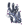
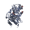
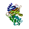
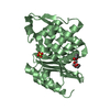
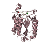
| Title | Crystal structure of serratia fonticola carbapenemase SFC-1 S70A-Meropenem complex | ||||||
 Components Components | Carbapenem-hydrolizing beta-lactamase SFC-1 | ||||||
 Keywords Keywords | HYDROLASE/Antibiotic / CARBAPENEMASE / HYDROLASE / ANTIBIOTIC RESISTANCE / BETA-LACTAMASE COMPLEX / HYDROLASE-Antibiotic complex | ||||||
| Function / homology |  Function and homology information Function and homology informationbeta-lactam antibiotic catabolic process / beta-lactamase activity / beta-lactamase / response to antibiotic Similarity search - Function | ||||||
| Biological species |  Serratia fonticola (bacteria) Serratia fonticola (bacteria) | ||||||
| Method |  X-RAY DIFFRACTION / SYNCHROTRON / MOLECULAR REPLACEMENT / Resolution: 1.08 Å X-RAY DIFFRACTION / SYNCHROTRON / MOLECULAR REPLACEMENT / Resolution: 1.08 Å | ||||||
 Authors Authors | Fonseca, F. / Spencer, J. | ||||||
 Citation Citation | Journal: J.Am.Chem.Soc. / Year: 2012 Title: The basis for carbapenem hydrolysis by class A beta-lactamases: a combined investigation using crystallography and simulations. Authors: Fonseca, F. / Chudyk, E.I. / van der Kamp, M.W. / Correia, A. / Mulholland, A.J. / Spencer, J. | ||||||
| History |
|
- Structure visualization
| Structure viewer | Molecule: MolmilJmol/JSmol |
|---|
- Downloads & links
-Download
| PDBx/mmCIF format | 4euz.cif.gz | 154.6 KB | Display | PDBx/mmCIF format |
|---|---|---|---|---|
| PDB format | pdb4euz.ent.gz | 118.5 KB | Display | PDB format |
| PDBx/mmJSON format | 4euz.json.gz | Tree view | PDBx/mmJSON format | |
| Others |  Other downloads Other downloads |
-Validation report
| Arichive directory | https://data.pdbj.org/pub/pdb/validation_reports/eu/4euzftp://data.pdbj.org/pub/pdb/validation_reports/eu/4euz | HTTPS FTP |
|---|
-Related structure data
| Related structure data | 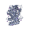 4eqiSC  4ev4C S: Starting model for refinement C: citing same article ( |
|---|---|
| Similar structure data |


-Links
 PDBj
PDBj


- Assembly
| Deposited unit | 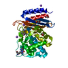
| ||||||||
|---|---|---|---|---|---|---|---|---|---|
| 1 |
| ||||||||
| Unit cell |
|
-Components
| #1: Protein | Mass: 30809.768 Da / Num. of mol.: 1 / Fragment: UNP residues 27-309 / Mutation: S70A Source method: isolated from a genetically manipulated source Source: (gene. exp.) Serratia fonticola (bacteria) / Strain: UTAD54 / Gene: BLASFC-1, SFC-1 / Plasmid: pET26-b / Production host: | ||||||
|---|---|---|---|---|---|---|---|
| #2: Chemical | ChemComp-MEM / (  Mass: 383.463 Da / Num. of mol.: 1 / Source method: obtained synthetically / Formula: C17H25N3O5S / Comment: antibiotic*YM Mass: 383.463 Da / Num. of mol.: 1 / Source method: obtained synthetically / Formula: C17H25N3O5S / Comment: antibiotic*YM | ||||||
| #3: Chemical | ChemComp-EDO /   Mass: 62.068 Da / Num. of mol.: 4 / Source method: obtained synthetically / Formula: C2H6O2 Mass: 62.068 Da / Num. of mol.: 4 / Source method: obtained synthetically / Formula: C2H6O2#4: Chemical | ChemComp-NA /   Mass: 22.990 Da / Num. of mol.: 7 / Source method: obtained synthetically / Formula: Na Mass: 22.990 Da / Num. of mol.: 7 / Source method: obtained synthetically / Formula: Na#5: Water | ChemComp-HOH / |  Mass: 18.015 Da / Num. of mol.: 494 / Source method: isolated from a natural source / Formula: H2O Mass: 18.015 Da / Num. of mol.: 494 / Source method: isolated from a natural source / Formula: H2OSequence details | THE TWO SEQADV RECORDS RELATED TO RESIDUES 255 AND 270 REPRESENT DISCREPANCIES BETWEEN THE ...THE TWO SEQADV RECORDS RELATED TO RESIDUES 255 AND 270 REPRESENT DISCREPANC | |
-Experimental details
-Experiment
| Experiment | Method: X-RAY DIFFRACTION / Number of used crystals: 1 |
|---|
- Sample preparation
| Crystal | Density Matthews: 2.34 Å3/Da / Density % sol: 47.53 % |
|---|---|
| Crystal grow | Temperature: 293 K / Method: vapor diffusion, hanging drop / pH: 7 Details: Na acetate, PEG 3350, pH 7, VAPOR DIFFUSION, HANGING DROP, temperature 293K |
-Data collection
| Diffraction | Mean temperature: 100 K | ||||||||||||
|---|---|---|---|---|---|---|---|---|---|---|---|---|---|
| Diffraction source | Source: SYNCHROTRON / Site: Diamond  / Beamline: I02 / Wavelength: 0.9702 Å / Beamline: I02 / Wavelength: 0.9702 Å | ||||||||||||
| Detector | Type: ADSC QUANTUM 315 / Detector: CCD / Date: Jul 21, 2009 | ||||||||||||
| Radiation | Monochromator: SI(111) DOUBLE CRYSTAL / Protocol: SINGLE WAVELENGTH / Monochromatic (M) / Laue (L): M / Scattering type: x-ray | ||||||||||||
| Radiation wavelength | Wavelength: 0.9702 Å / Relative weight: 1 | ||||||||||||
| Reflection | Resolution: 1.08→48.732 Å / Num. all: 123160 / Num. obs: 123160 / % possible obs: 99.5 % / Redundancy: 6.8 % / Biso Wilson estimate: 5.9 Å2 / Rmerge(I) obs: 0.07 / Net I/σ(I): 16.9 | ||||||||||||
| Reflection shell |
|
- Processing
| Software |
| |||||||||||||||||||||||||||||||||
|---|---|---|---|---|---|---|---|---|---|---|---|---|---|---|---|---|---|---|---|---|---|---|---|---|---|---|---|---|---|---|---|---|---|---|
| Refinement | Method to determine structure: MOLECULAR REPLACEMENT Starting model: PDB entry 4EQI Resolution: 1.08→22.42 Å / Num. parameters: 25608 / Num. restraintsaints: 33525 / Cross valid method: FREE R / σ(F): 0 / Stereochemistry target values: Engh & Huber Details: ANISOTROPIC REFINEMENT REDUCED FREE R (NO CUTOFF) BY 3.19%
| |||||||||||||||||||||||||||||||||
| Refine analyze | Num. disordered residues: 41 / Occupancy sum hydrogen: 1961.3 / Occupancy sum non hydrogen: 2603.05 | |||||||||||||||||||||||||||||||||
| Refinement step | Cycle: LAST / Resolution: 1.08→22.42 Å
| |||||||||||||||||||||||||||||||||
| Refine LS restraints |
|
