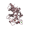
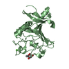
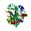
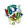
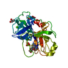
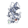
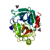
- PDB-4esq: Crystal structure of the extracellular domain of PknH from Mycoba... -
+
Open data
ID or keywords:
Loading...
-
Basic information
Entry
Database: PDB / ID: 4esq
Title
Crystal structure of the extracellular domain of PknH from Mycobacterium tuberculosis
Components
Serine/threonine protein kinase
Keywords
TRANSFERASE / Receptor Kinase / Membrane
Function / homology
Function and homology information
regulation of cell wall organization or biogenesis / regulation of primary metabolic process / negative regulation of growth / peptidoglycan-based cell wall / protein kinase activity / non-specific serine/threonine protein kinase / protein serine kinase activity / protein serine/threonine kinase activity / regulation of DNA-templated transcription / positive regulation of DNA-templated transcription ...regulation of cell wall organization or biogenesis / regulation of primary metabolic process / negative regulation of growth / peptidoglycan-based cell wall / protein kinase activity / non-specific serine/threonine protein kinase / protein serine kinase activity / protein serine/threonine kinase activity / regulation of DNA-templated transcription / positive regulation of DNA-templated transcription / ATP binding / plasma membrane Similarity search - Function
PknH-like extracellular domain / PknH-like extracellular domain / PknH-like extracellular domain superfamily / PknH-like extracellular domain / Protein Transport Mog1p; Chain A / Serine/threonine-protein kinase, active site / Serine/Threonine protein kinases active-site signature. / Protein kinase domain / Serine/Threonine protein kinases, catalytic domain / Protein kinase, ATP binding site ...PknH-like extracellular domain / PknH-like extracellular domain / PknH-like extracellular domain superfamily / PknH-like extracellular domain / Protein Transport Mog1p; Chain A / Serine/threonine-protein kinase, active site / Serine/Threonine protein kinases active-site signature. / Protein kinase domain / Serine/Threonine protein kinases, catalytic domain / Protein kinase, ATP binding site / Protein kinases ATP-binding region signature. / Protein kinase domain profile. / Protein kinase domain / Protein kinase-like domain superfamily / 3-Layer(aba) Sandwich / Alpha Beta Similarity search - Domain/homology
TERBIUM(III) ION / non-specific serine/threonine protein kinase / Serine/threonine-protein kinase PknH Similarity search - Component
Biological species
Mycobacterium tuberculosis (bacteria)
Method
X-RAY DIFFRACTION / SYNCHROTRON / SAD / Resolution: 1.7 Å
Monochromator: Double flat crystal, Si(111) / Protocol: SINGLE WAVELENGTH / Monochromatic (M) / Laue (L): M / Scattering type: x-ray
Radiation wavelength
Wavelength: 1.1159 Å / Relative weight: 1
Reflection
Redundancy: 3.8 % / Av σ(I) over netI: 17.44 / Number: 69365 / Rmerge(I) obs: 0.061 / Χ2: 1.01 / D res high: 1.7 Å / D res low: 50 Å / Num. obs: 18089 / % possible obs: 98.8
Diffraction reflection shell
Highest resolution (Å)
Lowest resolution (Å)
% possible obs (%)
ID
Rmerge(I) obs
Chi squared
Redundancy
3.66
50
99.7
1
0.04
0.98
3.8
2.91
3.66
99.9
1
0.055
0.96
3.8
2.54
2.91
99.7
1
0.061
1.081
3.9
2.31
2.54
99.6
1
0.068
1.026
3.9
2.14
2.31
99.1
1
0.073
1.077
3.9
2.02
2.14
98.9
1
0.086
1.064
3.9
1.91
2.02
98.2
1
0.108
1.01
3.9
1.83
1.91
97.7
1
0.148
1.005
3.8
1.76
1.83
97.5
1
0.198
1.017
3.8
1.7
1.76
98.1
1
0.253
0.927
3.8
Reflection
Resolution: 1.7→50 Å / Num. obs: 34200 / % possible obs: 98.8 % / Redundancy: 3.8 % / Rmerge(I) obs: 0.061 / Χ2: 1.015 / Net I/σ(I): 17.1
In the structure databanks used in Yorodumi, some data are registered as the other names, "COVID-19 virus" and "2019-nCoV". Here are the details of the virus and the list of structure data.
Jan 31, 2019. EMDB accession codes are about to change! (news from PDBe EMDB page)
EMDB accession codes are about to change! (news from PDBe EMDB page)
The allocation of 4 digits for EMDB accession codes will soon come to an end. Whilst these codes will remain in use, new EMDB accession codes will include an additional digit and will expand incrementally as the available range of codes is exhausted. The current 4-digit format prefixed with “EMD-” (i.e. EMD-XXXX) will advance to a 5-digit format (i.e. EMD-XXXXX), and so on. It is currently estimated that the 4-digit codes will be depleted around Spring 2019, at which point the 5-digit format will come into force.
The EM Navigator/Yorodumi systems omit the EMD- prefix.
Related info.:Q: What is EMD? / ID/Accession-code notation in Yorodumi/EM Navigator
Yorodumi is a browser for structure data from EMDB, PDB, SASBDB, etc.
This page is also the successor to EM Navigator detail page, and also detail information page/front-end page for Omokage search.
The word "yorodu" (or yorozu) is an old Japanese word meaning "ten thousand". "mi" (miru) is to see.
Related info.:EMDB / PDB / SASBDB / Comparison of 3 databanks / Yorodumi Search / Aug 31, 2016. New EM Navigator & Yorodumi / Yorodumi Papers / Jmol/JSmol / Function and homology information / Changes in new EM Navigator and Yorodumi
 Movie
Movie Controller

 Yorodumi
 Open data
 Basic information
 Components
 Keywords
 Function and homology information
 Mycobacterium tuberculosis (bacteria)
 X-RAY DIFFRACTION /
  Authors
 Citation
 Structure visualization
 Downloads & links
 Other downloads
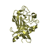


 PDBj
PDBj Assembly
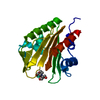

 Mass: 209.240 Da / Num. of mol.: 1 / Source method: obtained synthetically / Formula: C8H19NO5 / Comment: pH buffer*YM
 Mass: 158.925 Da / Num. of mol.: 2 / Source method: obtained synthetically / Formula: Tb
 Mass: 18.015 Da / Num. of mol.: 221 / Source method: isolated from a natural source / Formula: H2O
 Sample preparation
 / Beamline: 8.3.1 / Wavelength: 1.1159 Å
 Processing