 Movie
Movie Controller

[English] 日本語
 Yorodumi
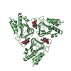
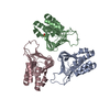
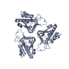
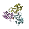
- PDB-4e6u: Structure of LpxA from Acinetobacter baumannii at 1.4A resolution... -
+ Open data

- Basic information
| Entry | Database: PDB / ID: 4e6u | ||||||
|---|---|---|---|---|---|---|---|
| Title | Structure of LpxA from Acinetobacter baumannii at 1.4A resolution (P63 form) | ||||||
 Components Components | Acyl-[acyl-carrier-protein]--UDP-N-acetylglucosamine O-acyltransferase | ||||||
 Keywords Keywords | TRANSFERASE / lipopolysaccaride synthesis | ||||||
| Function / homology |  Function and homology information Function and homology informationUdp N-acetylglucosamine O-acyltransferase; Domain 2 / Udp N-acetylglucosamine O-acyltransferase, C-terminal domain / Hexapeptide repeat proteins / UDP N-Acetylglucosamine Acyltransferase; domain 1 / 3 Solenoid / Up-down Bundle / Mainly Beta / Mainly Alpha Similarity search - Domain/homology | ||||||
| Biological species |  Acinetobacter baumannii (bacteria) Acinetobacter baumannii (bacteria) | ||||||
| Method |  X-RAY DIFFRACTION / SYNCHROTRON / 4E6T / Resolution: 1.41 Å X-RAY DIFFRACTION / SYNCHROTRON / 4E6T / Resolution: 1.41 Å | ||||||
 Authors Authors | Badger, J. / Chie-Leon, B. / Logan, C. / Sridhar, V. / Sankaran, B. / Zwart, P.H. / Nienaber, V. | ||||||
 Citation Citation | Journal: Acta Crystallogr.,Sect.F / Year: 2012 Title: Structure determination of LpxA from the lipopolysaccharide-synthesis pathway of Acinetobacter baumannii. Authors: Badger, J. / Chie-Leon, B. / Logan, C. / Sridhar, V. / Sankaran, B. / Zwart, P.H. / Nienaber, V. | ||||||
| History |
|
- Structure visualization
| Structure viewer | Molecule: MolmilJmol/JSmol |
|---|
- Downloads & links
-Download
| PDBx/mmCIF format | 4e6u.cif.gz | 71.3 KB | Display | PDBx/mmCIF format |
|---|---|---|---|---|
| PDB format | pdb4e6u.ent.gz | 52.4 KB | Display | PDB format |
| PDBx/mmJSON format | 4e6u.json.gz | Tree view | PDBx/mmJSON format | |
| Others |  Other downloads Other downloads |
-Validation report
| Arichive directory | https://data.pdbj.org/pub/pdb/validation_reports/e6/4e6uftp://data.pdbj.org/pub/pdb/validation_reports/e6/4e6u | HTTPS FTP |
|---|
-Related structure data


-Links
 PDBj
PDBj- Assembly
| Deposited unit | 
| |||||||||||||||||||||
|---|---|---|---|---|---|---|---|---|---|---|---|---|---|---|---|---|---|---|---|---|---|---|
| 1 | 
| |||||||||||||||||||||
| Unit cell |
| |||||||||||||||||||||
| Components on special symmetry positions |
|
-Components
| #1: Protein | Mass: 28472.334 Da / Num. of mol.: 1 Source method: isolated from a genetically manipulated source Source: (gene. exp.) Acinetobacter baumannii (bacteria) / Gene: lpxA, ABTW07_2294 / Cell line (production host): BL21 (DE3) / Production host: References: UniProt: F0QHB3, UniProt: A0A7U4DSW1*PLUS, acyl-[acyl-carrier-protein]-UDP-N-acetylglucosamine O-acyltransferase | ||||
|---|---|---|---|---|---|
| #2: Chemical |   Mass: 96.063 Da / Num. of mol.: 2 / Source method: obtained synthetically / Formula: SO4 Mass: 96.063 Da / Num. of mol.: 2 / Source method: obtained synthetically / Formula: SO4#3: Chemical | ChemComp-EDO /   Mass: 62.068 Da / Num. of mol.: 9 / Source method: obtained synthetically / Formula: C2H6O2 Mass: 62.068 Da / Num. of mol.: 9 / Source method: obtained synthetically / Formula: C2H6O2#4: Water | ChemComp-HOH / |  Mass: 18.015 Da / Num. of mol.: 293 / Source method: isolated from a natural source / Formula: H2O Mass: 18.015 Da / Num. of mol.: 293 / Source method: isolated from a natural source / Formula: H2O |
-Experimental details
-Experiment
| Experiment | Method: X-RAY DIFFRACTION / Number of used crystals: 1 |
|---|
- Sample preparation
| Crystal | Density Matthews: 3.46 Å3/Da / Density % sol: 64.49 % |
|---|---|
| Crystal grow | Temperature: 293 K / Method: vapor diffusion, hanging drop / pH: 7.5 Details: 2ul of 25mg/ml protein, 2uL of 1.8M LiSO4, 0.1M Hepes 7.5, VAPOR DIFFUSION, HANGING DROP, temperature 293K |
-Data collection
| Diffraction | Mean temperature: 100 K |
|---|---|
| Diffraction source | Source: SYNCHROTRON / Site: APS  / Beamline: 21-ID-F / Wavelength: 0.979 Å / Beamline: 21-ID-F / Wavelength: 0.979 Å |
| Detector | Type: RAYONIX MX-225 / Detector: CCD / Date: Nov 6, 2010 |
| Radiation | Protocol: SINGLE WAVELENGTH / Monochromatic (M) / Laue (L): M / Scattering type: x-ray |
| Radiation wavelength | Wavelength: 0.979 Å / Relative weight: 1 |
| Reflection | Resolution: 1.41→50 Å / Num. all: 73881 / Num. obs: 73881 / % possible obs: 99.8 % / Observed criterion σ(I): -4 / Redundancy: 11.3 % / Rmerge(I) obs: 0.064 / Net I/σ(I): 36.1 |
| Reflection shell | Resolution: 1.41→1.46 Å / Redundancy: 11.2 % / Rmerge(I) obs: 0.591 / Mean I/σ(I) obs: 5 / Num. unique all: 63291 / % possible all: 99.8 |
- Processing
| Software |
| |||||||||||||||||||||||||||||||||||||||||||||||||||||||||||||||||
|---|---|---|---|---|---|---|---|---|---|---|---|---|---|---|---|---|---|---|---|---|---|---|---|---|---|---|---|---|---|---|---|---|---|---|---|---|---|---|---|---|---|---|---|---|---|---|---|---|---|---|---|---|---|---|---|---|---|---|---|---|---|---|---|---|---|---|
| Refinement | Method to determine structure: 4E6T / Resolution: 1.41→36.09 Å / Cor.coef. Fo:Fc: 0.97 / Cor.coef. Fo:Fc free: 0.968 / SU B: 0.758 / SU ML: 0.031 / Cross valid method: THROUGHOUT / ESU R: 0.049 / ESU R Free: 0.05 / Stereochemistry target values: MAXIMUM LIKELIHOOD
| |||||||||||||||||||||||||||||||||||||||||||||||||||||||||||||||||
| Solvent computation | Ion probe radii: 0.8 Å / Shrinkage radii: 0.8 Å / VDW probe radii: 1.4 Å / Solvent model: MASK | |||||||||||||||||||||||||||||||||||||||||||||||||||||||||||||||||
| Displacement parameters | Biso mean: 20.799 Å2
| |||||||||||||||||||||||||||||||||||||||||||||||||||||||||||||||||
| Refinement step | Cycle: LAST / Resolution: 1.41→36.09 Å
| |||||||||||||||||||||||||||||||||||||||||||||||||||||||||||||||||
| Refine LS restraints |
| |||||||||||||||||||||||||||||||||||||||||||||||||||||||||||||||||
| LS refinement shell | Resolution: 1.412→1.449 Å / Total num. of bins used: 20
|
