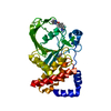
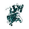
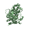
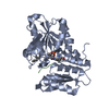
- PDB-4e6e: Crystal structure of a putative cell division protein FtsZ (Tfu_1... -
+
Open data
ID or keywords:
Loading...
-
Basic information
Entry
Database: PDB / ID: 4e6e
Title
Crystal structure of a putative cell division protein FtsZ (Tfu_1113) from Thermobifida fusca YX-ER1 at 2.22 A resolution (PSI Community Target, van Wezel G.P.)
Components
Cell division protein ftsZ
Keywords
CELL CYCLE / Cell division / tubulin homolog / GTP-binding / Structural Genomics / Joint Center for Structural Genomics / JCSG / Protein Structure Initiative / PSI-BIOLOGY
Function / homology
Function and homology information
division septum assembly / FtsZ-dependent cytokinesis / cell division site / protein polymerization / GTPase activity / GTP binding / metal ion binding / cytoplasm Similarity search - Function
FtsZ protein signature 2. / Cell division protein FtsZ / Cell division protein FtsZ, conserved site / Cell division protein FtsZ, C-terminal / FtsZ family, C-terminal domain / FtsZ protein signature 1. / Tubulin-like protein FtsZ/CetZ / Tubulin/FtsZ, C-terminal domain / Tubulin/FtsZ, GTPase domain / 60s Ribosomal Protein L30; Chain: A; ...FtsZ protein signature 2. / Cell division protein FtsZ / Cell division protein FtsZ, conserved site / Cell division protein FtsZ, C-terminal / FtsZ family, C-terminal domain / FtsZ protein signature 1. / Tubulin-like protein FtsZ/CetZ / Tubulin/FtsZ, C-terminal domain / Tubulin/FtsZ, GTPase domain / 60s Ribosomal Protein L30; Chain: A; / Tubulin/FtsZ family, C-terminal domain / Tubulin/FtsZ-like, C-terminal domain / Tubulin/FtsZ, C-terminal / Tubulin/FtsZ, 2-layer sandwich domain / Tubulin/FtsZ family, GTPase domain / Tubulin/FtsZ family, GTPase domain / Tubulin/FtsZ, GTPase domain / Tubulin/FtsZ, GTPase domain superfamily / Rossmann fold / 2-Layer Sandwich / 3-Layer(aba) Sandwich / Alpha Beta Similarity search - Domain/homology
Mass: 18.015 Da / Num. of mol.: 139 / Source method: isolated from a natural source / Formula: H2O
Has protein modification
Y
Sequence details
1. THE CONSTRUCT (1-313) WAS EXPRESSED WITH AN N-TERMINAL PURIFICATION TAG MGSDKIHHHHHHENLYFQG. THE ...1. THE CONSTRUCT (1-313) WAS EXPRESSED WITH AN N-TERMINAL PURIFICATION TAG MGSDKIHHHHHHENLYFQG. THE TAG WAS REMOVED WITH TEV PROTEASE LEAVING ONLY A GLYCINE (0) FOLLOWED BY THE TARGET SEQUENCE. 2. THIS GENE USES AN ALTERNATE INITIATION CODON THAT RESULTS IN A VALINE AT POSITION 1 WHEN EXPRESSED AS A FUSION WITH THE PURIFICATION TAG.
-
Experimental details
-
Experiment
Experiment
Method: X-RAY DIFFRACTION / Number of used crystals: 1
-
Sample preparation
Crystal
Density Matthews: 2.69 Å3/Da / Density % sol: 54.21 %
Crystal grow
Temperature: 293 K / Method: vapor diffusion, sitting drop / pH: 6.5 Details: 1.600M magnesium sulfate, 0.1M MES pH 6.5, NANODROP, VAPOR DIFFUSION, SITTING DROP, temperature 293K
Method to determine structure: SAD / Resolution: 2.12→28.226 Å / Cor.coef. Fo:Fc: 0.9561 / Cor.coef. Fo:Fc free: 0.9311 / Occupancy max: 1 / Occupancy min: 0.5 / Cross valid method: THROUGHOUT / σ(F): 0 Details: 1. A MET-INHIBITION PROTOCOL WAS USED FOR SELENOMETHIONINE INCORPORATION DURING PROTEIN EXPRESSION. THE OCCUPANCY OF THE SE ATOMS IN THE MSE RESIDUES WAS REDUCED TO 0.75 FOR THE REDUCED ...Details: 1. A MET-INHIBITION PROTOCOL WAS USED FOR SELENOMETHIONINE INCORPORATION DURING PROTEIN EXPRESSION. THE OCCUPANCY OF THE SE ATOMS IN THE MSE RESIDUES WAS REDUCED TO 0.75 FOR THE REDUCED SCATTERING POWER DUE TO PARTIAL S-MET INCORPORATION. 2. SULFATE (SO4), MAGNESIUM (MG) FROM CRYSTALLIZATION CONDITION AND CHLORIDE (CL) FROM THE PROTEIN BUFFER ARE MODELED INTO THE STRUCTURE 3. ATOM RECORD CONTAINS SUM OF TLS AND RESIDUAL B FACTORS. ANISOU RECORD CONTAINS SUM OF TLS AND RESIDUAL U FACTORS. 4. WATERS WERE EXCLUDED FROM AUTOMATIC TLS ASSIGNMENT. 5. SAD EXPERIMENTAL PHASES WERE USED AS RESTRAINTS DURING REFINEMENT.
In the structure databanks used in Yorodumi, some data are registered as the other names, "COVID-19 virus" and "2019-nCoV". Here are the details of the virus and the list of structure data.
Jan 31, 2019. EMDB accession codes are about to change! (news from PDBe EMDB page)
EMDB accession codes are about to change! (news from PDBe EMDB page)
The allocation of 4 digits for EMDB accession codes will soon come to an end. Whilst these codes will remain in use, new EMDB accession codes will include an additional digit and will expand incrementally as the available range of codes is exhausted. The current 4-digit format prefixed with “EMD-” (i.e. EMD-XXXX) will advance to a 5-digit format (i.e. EMD-XXXXX), and so on. It is currently estimated that the 4-digit codes will be depleted around Spring 2019, at which point the 5-digit format will come into force.
The EM Navigator/Yorodumi systems omit the EMD- prefix.
Related info.:Q: What is EMD? / ID/Accession-code notation in Yorodumi/EM Navigator
Yorodumi is a browser for structure data from EMDB, PDB, SASBDB, etc.
This page is also the successor to EM Navigator detail page, and also detail information page/front-end page for Omokage search.
The word "yorodu" (or yorozu) is an old Japanese word meaning "ten thousand". "mi" (miru) is to see.
Related info.:EMDB / PDB / SASBDB / Comparison of 3 databanks / Yorodumi Search / Aug 31, 2016. New EM Navigator & Yorodumi / Yorodumi Papers / Jmol/JSmol / Function and homology information / Changes in new EM Navigator and Yorodumi
 Movie
Movie Controller

 Yorodumi
 Open data
 Basic information
 Components
 Keywords
 Function and homology information
 Thermobifida fusca (bacteria)
 X-RAY DIFFRACTION /
  Authors
 Citation
 Structure visualization
 Downloads & links
 Other downloads
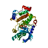


 PDBj
PDBj


 Assembly
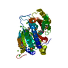


 Mass: 24.305 Da / Num. of mol.: 4 / Source method: obtained synthetically / Formula: Mg
 Mass: 35.453 Da / Num. of mol.: 2 / Source method: obtained synthetically / Formula: Cl
 Mass: 96.063 Da / Num. of mol.: 1 / Source method: obtained synthetically / Formula: SO4
 Mass: 18.015 Da / Num. of mol.: 139 / Source method: isolated from a natural source / Formula: H2O
 Sample preparation
 / Beamline: BL11-1 / Wavelength: 0.97895
  Processing