 Movie
Movie Controller


+ Open data

- Basic information
| Entry | Database: PDB / ID: 4.0E+58 | ||||||
|---|---|---|---|---|---|---|---|
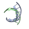
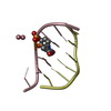
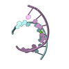
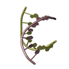
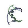
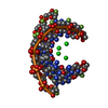
| Title | Crystal structure of GCC(LCG)CCGC duplex containing LNA residue | ||||||
 Components Components | RNA duplex containing CCG repeats | ||||||
 Keywords Keywords | RNA / CCG repeats / 5' slippery duplexes / X-linked mental retardation / Huntington's disease / myotonic dystrophy type 1 / LNA Guanosine | ||||||
| Function / homology | RNA Function and homology information Function and homology information | ||||||
| Biological species |  Homo sapiens (human) Homo sapiens (human) | ||||||
| Method |  X-RAY DIFFRACTION / SYNCHROTRON / MOLECULAR REPLACEMENT / Resolution: 1.952 Å X-RAY DIFFRACTION / SYNCHROTRON / MOLECULAR REPLACEMENT / Resolution: 1.952 Å | ||||||
 Authors Authors | Kiliszek, A. / Kierzek, R. / Krzyzosiak, W.J. / Rypniewski, R. | ||||||
 Citation Citation | Journal: Nucleic Acids Res. / Year: 2012 Title: Crystallographic characterization of CCG repeats. Authors: Kiliszek, A. / Kierzek, R. / Krzyzosiak, W.J. / Rypniewski, W. | ||||||
| History |
|
- Structure visualization
| Structure viewer | Molecule: MolmilJmol/JSmol |
|---|
- Downloads & links
-Download
| PDBx/mmCIF format | 4e58.cif.gz | 21.9 KB | Display | PDBx/mmCIF format |
|---|---|---|---|---|
| PDB format | pdb4e58.ent.gz | 15.2 KB | Display | PDB format |
| PDBx/mmJSON format | 4e58.json.gz | Tree view | PDBx/mmJSON format | |
| Others |  Other downloads Other downloads |
-Validation report
| Arichive directory | https://data.pdbj.org/pub/pdb/validation_reports/e5/4e58ftp://data.pdbj.org/pub/pdb/validation_reports/e5/4e58 | HTTPS FTP |
|---|
-Related structure data
| Related structure data | 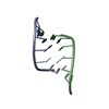 4e59C 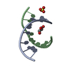 3glpS C: citing same article ( S: Starting model for refinement |
|---|---|
| Similar structure data |


-Links
 PDBj
PDBj- Assembly
| Deposited unit | 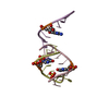
| ||||||||
|---|---|---|---|---|---|---|---|---|---|
| 1 | 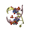
| ||||||||
| 2 | 
| ||||||||
| Unit cell |
| ||||||||
| Components on special symmetry positions |
|
-Components
| #1: RNA chain | Mass: 2528.579 Da / Num. of mol.: 3 / Source method: obtained synthetically / Details: Similar sequences occur in mRNA in humans / Source: (synth.) Homo sapiens (human)#2: Chemical |   Mass: 96.063 Da / Num. of mol.: 2 / Source method: obtained synthetically / Formula: SO4 Mass: 96.063 Da / Num. of mol.: 2 / Source method: obtained synthetically / Formula: SO4#3: Water | ChemComp-HOH / |  Mass: 18.015 Da / Num. of mol.: 14 / Source method: isolated from a natural source / Formula: H2O Mass: 18.015 Da / Num. of mol.: 14 / Source method: isolated from a natural source / Formula: H2O |
|---|
-Experimental details
-Experiment
| Experiment | Method: X-RAY DIFFRACTION / Number of used crystals: 1 |
|---|
- Sample preparation
| Crystal | Density Matthews: 2.38 Å3/Da / Density % sol: 48.22 % |
|---|---|
| Crystal grow | Temperature: 292 K / Method: vapor diffusion, sitting drop / pH: 5.6 Details: MES, MgCl2 and Li2SO4, pH 5.6, VAPOR DIFFUSION, SITTING DROP, temperature 292.0K |
-Data collection
| Diffraction | Mean temperature: 100 K |
|---|---|
| Diffraction source | Source: SYNCHROTRON / Site: BESSY  / Beamline: 14.1 / Wavelength: 0.9148 Å / Beamline: 14.1 / Wavelength: 0.9148 Å |
| Detector | Type: MARMOSAIC 225 mm CCD / Detector: CCD / Date: Mar 19, 2011 |
| Radiation | Monochromator: Si / Protocol: SINGLE WAVELENGTH / Monochromatic (M) / Laue (L): M / Scattering type: x-ray |
| Radiation wavelength | Wavelength: 0.9148 Å / Relative weight: 1 |
| Reflection | Resolution: 1.95→20 Å / Num. all: 5762 / Num. obs: 5762 / % possible obs: 99.8 % / Observed criterion σ(F): 0 / Observed criterion σ(I): 0 / Redundancy: 28.3 % / Biso Wilson estimate: 50.7 Å2 / Rsym value: 0.067 / Net I/σ(I): 23.7 |
| Reflection shell | Resolution: 1.95→1.98 Å / Redundancy: 15.6 % / Mean I/σ(I) obs: 2.175 / Num. unique all: 273 / Rsym value: 0.673 / % possible all: 97.5 |
- Processing
| Software |
| |||||||||||||||||||||||||
|---|---|---|---|---|---|---|---|---|---|---|---|---|---|---|---|---|---|---|---|---|---|---|---|---|---|---|
| Refinement | Method to determine structure: MOLECULAR REPLACEMENT Starting model: PDB ENTRY 3GLP Resolution: 1.952→18.79 Å / SU ML: 0.36 / Isotropic thermal model: Isotropic / Cross valid method: THROUGHOUT / σ(F): 0 / Phase error: 31.83 / Stereochemistry target values: ML
| |||||||||||||||||||||||||
| Solvent computation | Shrinkage radii: 0.49 Å / VDW probe radii: 0.8 Å / Solvent model: FLAT BULK SOLVENT MODEL / Bsol: 36.22 Å2 / ksol: 0.348 e/Å3 | |||||||||||||||||||||||||
| Displacement parameters | Biso mean: 49.43 Å2
| |||||||||||||||||||||||||
| Refinement step | Cycle: LAST / Resolution: 1.952→18.79 Å
| |||||||||||||||||||||||||
| Refine LS restraints |
| |||||||||||||||||||||||||
| LS refinement shell | Resolution: 1.952→2.1486 Å
|
