 Movie
Movie Controller

[English] 日本語
 Yorodumi
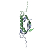
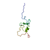
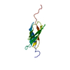
- PDB-4e1p: Crystal structure of the dimerization domain of Lsr2 from Mycobac... -
+ Open data

- Basic information
| Entry | Database: PDB / ID: 4e1p | ||||||
|---|---|---|---|---|---|---|---|
| Title | Crystal structure of the dimerization domain of Lsr2 from Mycobacterium tuberculosis in the P 1 21 1 space group | ||||||
 Components Components | Protein lsr2 | ||||||
 Keywords Keywords | DNA BINDING PROTEIN / anti-parallel beta sheet / dimer | ||||||
| Function / homology |  Function and homology information Function and homology informationDNA protection / cellular response to oxygen levels / nucleoid / response to iron ion / acyltransferase activity / peptidoglycan-based cell wall / response to hydrogen peroxide / regulation of DNA-templated transcription / DNA binding / plasma membrane ...DNA protection / cellular response to oxygen levels / nucleoid / response to iron ion / acyltransferase activity / peptidoglycan-based cell wall / response to hydrogen peroxide / regulation of DNA-templated transcription / DNA binding / plasma membrane / cytosol / cytoplasm Similarity search - Function | ||||||
| Biological species |   Mycobacterium tuberculosis (bacteria) Mycobacterium tuberculosis (bacteria) | ||||||
| Method |  X-RAY DIFFRACTION / AB INITIO PHASING / Resolution: 1.728 Å X-RAY DIFFRACTION / AB INITIO PHASING / Resolution: 1.728 Å | ||||||
 Authors Authors | Summers, E.L. / Meindl, K. / Uson, I. / Arcus, V.L. | ||||||
 Citation Citation | Journal: Plos One / Year: 2012 Title: The structure of the oligomerization domain of Lsr2 from Mycobacterium tuberculosis reveals a mechanism for chromosome organization and protection. Authors: Summers, E.L. / Meindl, K. / Uson, I. / Mitra, A.K. / Radjainia, M. / Colangeli, R. / Alland, D. / Arcus, V.L. | ||||||
| History |
|
- Structure visualization
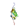
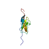
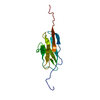
| Structure viewer | Molecule: MolmilJmol/JSmol |
|---|
- Downloads & links
-Download
| PDBx/mmCIF format | 4e1p.cif.gz | 36.4 KB | Display | PDBx/mmCIF format |
|---|---|---|---|---|
| PDB format | pdb4e1p.ent.gz | 25 KB | Display | PDB format |
| PDBx/mmJSON format | 4e1p.json.gz | Tree view | PDBx/mmJSON format | |
| Others |  Other downloads Other downloads |
-Validation report
| Arichive directory | https://data.pdbj.org/pub/pdb/validation_reports/e1/4e1pftp://data.pdbj.org/pub/pdb/validation_reports/e1/4e1p | HTTPS FTP |
|---|
-Related structure data


-Links
 PDBj
PDBj

- Assembly
| Deposited unit | 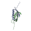
| ||||||||
|---|---|---|---|---|---|---|---|---|---|
| 1 |
| ||||||||
| Unit cell |
|
-Components
| #1: Protein | Mass: 6539.324 Da / Num. of mol.: 2 / Fragment: dimerization domain (UNP residues 1-61) Source method: isolated from a genetically manipulated source Source: (gene. exp.) Mycobacterium tuberculosis (bacteria) / Strain: H37Ra / Gene: lsr2, MT3704, MTCY07H7B.25, Rv3597c / Plasmid: pPROExHTb / Production host: #2: Water | ChemComp-HOH / |  Mass: 18.015 Da / Num. of mol.: 206 / Source method: isolated from a natural source / Formula: H2O Mass: 18.015 Da / Num. of mol.: 206 / Source method: isolated from a natural source / Formula: H2O |
|---|
-Experimental details
-Experiment
| Experiment | Method: X-RAY DIFFRACTION / Number of used crystals: 1 |
|---|
- Sample preparation
| Crystal | Density Matthews: 1.9 Å3/Da / Density % sol: 35.39 % |
|---|---|
| Crystal grow | Temperature: 291 K / Method: vapor diffusion, hanging drop / pH: 8.5 Details: 26% PEG400, 0.12 M ammonium sulfate, 0.1 M Tris, pH 8.5, VAPOR DIFFUSION, HANGING DROP, temperature 291K |
-Data collection
| Diffraction | Mean temperature: 110 K |
|---|---|
| Diffraction source | Source: ROTATING ANODE / Type: RIGAKU RUH3R / Wavelength: 1.54179 Å |
| Detector | Type: MAR scanner 345 mm plate / Detector: IMAGE PLATE / Date: Sep 16, 2011 / Details: msc osmic optics |
| Radiation | Monochromator: Ni filter / Protocol: SINGLE WAVELENGTH / Monochromatic (M) / Laue (L): M / Scattering type: x-ray |
| Radiation wavelength | Wavelength: 1.54179 Å / Relative weight: 1 |
| Reflection | Resolution: 1.728→32.4 Å / Num. obs: 10538 / % possible obs: 99.4 % / Redundancy: 9.8 % / Rmerge(I) obs: 0.092 / Net I/σ(I): 17.1 |
| Reflection shell | Resolution: 1.728→1.82 Å / Redundancy: 9.2 % / Rmerge(I) obs: 0.308 / Mean I/σ(I) obs: 6.9 / % possible all: 96.3 |
- Processing
| Software |
| |||||||||||||||||||||||||||||||||||
|---|---|---|---|---|---|---|---|---|---|---|---|---|---|---|---|---|---|---|---|---|---|---|---|---|---|---|---|---|---|---|---|---|---|---|---|---|
| Refinement | Method to determine structure: AB INITIO PHASING / Resolution: 1.728→29.097 Å / Occupancy max: 1 / Occupancy min: 1 / FOM work R set: 0.8996 / SU ML: 0.25 / σ(F): 0 / Phase error: 16.67 / Stereochemistry target values: ML
| |||||||||||||||||||||||||||||||||||
| Solvent computation | Shrinkage radii: 0.73 Å / VDW probe radii: 1 Å / Solvent model: FLAT BULK SOLVENT MODEL / Bsol: 41.831 Å2 / ksol: 0.382 e/Å3 | |||||||||||||||||||||||||||||||||||
| Displacement parameters | Biso max: 39.39 Å2 / Biso mean: 13.575 Å2 / Biso min: 3.37 Å2
| |||||||||||||||||||||||||||||||||||
| Refinement step | Cycle: LAST / Resolution: 1.728→29.097 Å
| |||||||||||||||||||||||||||||||||||
| Refine LS restraints |
| |||||||||||||||||||||||||||||||||||
| LS refinement shell | Refine-ID: X-RAY DIFFRACTION / Total num. of bins used: 4
|
