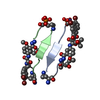
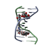
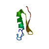
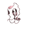
PROTEIN FIBRIL / amyloid / out-of-register / fiber-forming / macrocycle
Function / homology
Function and homology information
plus-end-directed organelle transport along microtubule / histone-dependent DNA binding / negative regulation of protein localization to mitochondrion / neurofibrillary tangle / microtubule lateral binding / axonal transport / tubulin complex / positive regulation of protein localization to synapse / phosphatidylinositol bisphosphate binding / generation of neurons ...plus-end-directed organelle transport along microtubule / histone-dependent DNA binding / negative regulation of protein localization to mitochondrion / neurofibrillary tangle / microtubule lateral binding / axonal transport / tubulin complex / positive regulation of protein localization to synapse / phosphatidylinositol bisphosphate binding / generation of neurons / rRNA metabolic process / axonal transport of mitochondrion / regulation of mitochondrial fission / axon development / regulation of microtubule-based movement / intracellular distribution of mitochondria / regulation of chromosome organization / central nervous system neuron development / minor groove of adenine-thymine-rich DNA binding / lipoprotein particle binding / microtubule polymerization / negative regulation of mitochondrial membrane potential / regulation of microtubule polymerization / dynactin binding / apolipoprotein binding / protein polymerization / main axon / Caspase-mediated cleavage of cytoskeletal proteins / regulation of microtubule polymerization or depolymerization / negative regulation of mitochondrial fission / axolemma / glial cell projection / neurofibrillary tangle assembly / positive regulation of axon extension / regulation of cellular response to heat / positive regulation of microtubule polymerization / positive regulation of protein localization / Activation of AMPK downstream of NMDARs / positive regulation of superoxide anion generation / cellular response to brain-derived neurotrophic factor stimulus / regulation of long-term synaptic depression / supramolecular fiber organization / cytoplasmic microtubule organization / regulation of calcium-mediated signaling / axon cytoplasm / somatodendritic compartment / synapse assembly / phosphatidylinositol binding / nuclear periphery / astrocyte activation / enzyme inhibitor activity / protein phosphatase 2A binding / stress granule assembly / regulation of microtubule cytoskeleton organization / regulation of autophagy / cellular response to reactive oxygen species / microglial cell activation / cellular response to nerve growth factor stimulus / Hsp90 protein binding / protein homooligomerization / SH3 domain binding / PKR-mediated signaling / synapse organization / regulation of synaptic plasticity / response to lead ion / microtubule cytoskeleton organization / memory / neuron projection development / cytoplasmic ribonucleoprotein granule / cell-cell signaling / single-stranded DNA binding / cellular response to heat / growth cone / protein-folding chaperone binding / microtubule cytoskeleton / actin binding / cell body / double-stranded DNA binding / sequence-specific DNA binding / microtubule binding / amyloid fibril formation / dendritic spine / microtubule / protein-macromolecule adaptor activity / learning or memory / neuron projection / membrane raft / negative regulation of gene expression / axon / neuronal cell body / DNA damage response / dendrite / protein kinase binding / enzyme binding / mitochondrion / DNA binding / RNA binding / extracellular region / identical protein binding / nucleus Similarity search - Function
Microtubule-associated protein Tau / Microtubule associated protein, tubulin-binding repeat / Tau and MAP protein, tubulin-binding repeat / Tau and MAP proteins tubulin-binding repeat signature. / Tau and MAP proteins tubulin-binding repeat profile. / : Similarity search - Domain/homology
Protocol: SINGLE WAVELENGTH / Monochromatic (M) / Laue (L): M / Scattering type: x-ray
Radiation wavelength
Wavelength: 0.91609 Å / Relative weight: 1
Reflection
Resolution: 1.75→18.22 Å / % possible obs: 99 % / Observed criterion σ(I): -3 / Biso Wilson estimate: 15.82 Å2 / Rmerge(I) obs: 0.086 / Net I/σ(I): 7.83
Reflection shell
Resolution (Å)
Highest resolution (Å)
Rmerge(I) obs
Mean I/σ(I) obs
Diffraction-ID
% possible all
1.75-1.8
0.424
2.5
1
98.7
1.8-1.84
0.414
2.55
1
98.8
1.84-1.9
0.286
3.52
1
99.7
1.9-1.96
0.225
4.23
1
99.3
1.96-2.02
0.162
4.99
1
99.5
2.02-2.09
0.138
6.15
1
99.4
2.09-2.17
0.125
7.11
1
99.5
2.17-2.26
0.132
7.12
1
99.3
2.26-2.36
0.118
7.84
1
100
2.36-2.48
0.108
8.02
1
98.4
2.48-2.61
0.109
8.8
1
99.8
2.61-2.77
0.083
10.4
1
99.3
2.77-2.96
0.085
10.77
1
99.5
2.96-3.2
0.068
12.43
1
98.4
3.2-3.5
0.058
14.11
1
100
3.5-3.91
0.058
15.04
1
98.6
3.91-4.52
0.056
15.53
1
97.3
4.52-5.53
0.041
16.41
1
97.2
5.53-7.83
0.052
15.66
1
95
7.83
0.044
14.97
1
89.4
-
Processing
Software
Name
Version
Classification
NB
XSCALE
datascaling
REFMAC
refinement
PDB_EXTRACT
3.1
dataextraction
BUSTER
2.10.0
refinement
Refinement
Method to determine structure: SAD / Resolution: 1.75→18.22 Å / Cor.coef. Fo:Fc: 0.9309 / Cor.coef. Fo:Fc free: 0.9272 / Occupancy max: 1 / Occupancy min: 0.5 / SU B: 4.937 / SU ML: 0.075 / SU R Cruickshank DPI: 0.1287 / Cross valid method: THROUGHOUT / σ(F): 0 / ESU R: 0.129 / ESU R Free: 0.109 / Stereochemistry target values: MAXIMUM LIKELIHOOD Details: HYDROGENS HAVE BEEN ADDED IN THE RIDING POSITIONS; U VALUES: RESIDUAL ONLY
Rfactor
Num. reflection
% reflection
Selection details
Rfree
0.2213
642
9.85 %
RANDOM
Rwork
0.1854
-
-
-
obs
0.1889
6519
99.86 %
-
Solvent computation
Ion probe radii: 0.8 Å / Shrinkage radii: 0.8 Å / VDW probe radii: 1.4 Å / Solvent model: MASK
Displacement parameters
Biso mean: 19.05 Å2
Baniso -1
Baniso -2
Baniso -3
1-
-2.8619 Å2
0 Å2
-3.8144 Å2
2-
-
1.8581 Å2
0 Å2
3-
-
-
1.0038 Å2
Refine analyze
Luzzati coordinate error obs: 0.18 Å
Refinement step
Cycle: LAST / Resolution: 1.75→18.22 Å
Protein
Nucleic acid
Ligand
Solvent
Total
Num. atoms
512
0
51
28
591
Refine LS restraints
Refine-ID
Type
Dev ideal
Number
Restraint function
Weight
X-RAY DIFFRACTION
t_bond_d
0.01
579
HARMONIC
2
X-RAY DIFFRACTION
t_angle_deg
1.29
703
HARMONIC
2
X-RAY DIFFRACTION
t_dihedral_angle_d
163
SINUSOIDAL
2
X-RAY DIFFRACTION
t_incorr_chiral_ct
X-RAY DIFFRACTION
t_pseud_angle
X-RAY DIFFRACTION
t_trig_c_planes
6
HARMONIC
2
X-RAY DIFFRACTION
t_gen_planes
55
HARMONIC
5
X-RAY DIFFRACTION
t_it
579
HARMONIC
20
X-RAY DIFFRACTION
t_nbd
X-RAY DIFFRACTION
t_omega_torsion
X-RAY DIFFRACTION
t_other_torsion
X-RAY DIFFRACTION
t_improper_torsion
X-RAY DIFFRACTION
t_chiral_improper_torsion
X-RAY DIFFRACTION
t_sum_occupancies
X-RAY DIFFRACTION
t_utility_distance
X-RAY DIFFRACTION
t_utility_angle
X-RAY DIFFRACTION
t_utility_torsion
X-RAY DIFFRACTION
t_ideal_dist_contact
LS refinement shell
Resolution: 1.75→1.96 Å / Total num. of bins used: 5
Rfactor
Num. reflection
% reflection
Rfree
0.2276
182
9.9 %
Rwork
0.1539
1657
-
all
0.1611
1839
-
obs
-
-
99.86 %
Refinement TLS params.
Method: refined / Refine-ID: X-RAY DIFFRACTION
ID
L11 (°2)
L12 (°2)
L13 (°2)
L22 (°2)
L23 (°2)
L33 (°2)
S11 (Å °)
S12 (Å °)
S13 (Å °)
S21 (Å °)
S22 (Å °)
S23 (Å °)
S31 (Å °)
S32 (Å °)
S33 (Å °)
T11 (Å2)
T12 (Å2)
T13 (Å2)
T22 (Å2)
T23 (Å2)
T33 (Å2)
Origin x (Å)
Origin y (Å)
Origin z (Å)
1
0.0753
1.6271
1.2704
1.745
0.3163
1.8355
0.0447
0.0558
0.1607
0.147
-0.0087
0.1412
0.0567
-0.004
-0.036
0.011
0.013
0.0192
-0.0115
-0.0117
-0.0023
-19.8576
-27.2394
-1.3919
2
0.9242
-1.227
0.0446
7.2824
2.6351
1.7875
0.1101
-0.0107
0.0987
-0.1211
-0.1104
0.0506
-0.1906
-0.1136
0.0003
-0.05
0.0137
0.0318
-0.0595
-0.0119
-0.0751
-19.1998
-17.7555
-3.2241
3
1.7149
0.513
0.3475
3.6902
0.6135
2.1247
0.0978
-0.1482
0.1472
0.0222
-0.1152
-0.0704
-0.0021
0.2262
0.0175
-0.1457
-0.0107
0.0268
-0.1138
0.0154
-0.0906
1.9368
-16.6877
-16.4196
4
1.391
-0.1185
-0.1887
4.5354
0.2754
1.469
-0.0514
-0.2411
-0.007
-0.0603
-0.0392
-0.1422
0.2287
0.0704
0.0906
-0.0646
-0.0038
0.0284
-0.03
0.015
-0.0582
2.8193
-26.4748
-17.7993
Refinement TLS group
ID
Refine-ID
Refine TLS-ID
Auth asym-ID
Auth seq-ID
1
X-RAY DIFFRACTION
1
A
0 - 13
2
X-RAY DIFFRACTION
2
B
0 - 13
3
X-RAY DIFFRACTION
3
C
0 - 13
4
X-RAY DIFFRACTION
4
D
0 - 13
+
About Yorodumi
-
News
-
Feb 9, 2022. New format data for meta-information of EMDB entries
New format data for meta-information of EMDB entries
Version 3 of the EMDB header file is now the official format.
The previous official version 1.9 will be removed from the archive.
In the structure databanks used in Yorodumi, some data are registered as the other names, "COVID-19 virus" and "2019-nCoV". Here are the details of the virus and the list of structure data.
Jan 31, 2019. EMDB accession codes are about to change! (news from PDBe EMDB page)
EMDB accession codes are about to change! (news from PDBe EMDB page)
The allocation of 4 digits for EMDB accession codes will soon come to an end. Whilst these codes will remain in use, new EMDB accession codes will include an additional digit and will expand incrementally as the available range of codes is exhausted. The current 4-digit format prefixed with “EMD-” (i.e. EMD-XXXX) will advance to a 5-digit format (i.e. EMD-XXXXX), and so on. It is currently estimated that the 4-digit codes will be depleted around Spring 2019, at which point the 5-digit format will come into force.
The EM Navigator/Yorodumi systems omit the EMD- prefix.
Related info.:Q: What is EMD? / ID/Accession-code notation in Yorodumi/EM Navigator
Yorodumi is a browser for structure data from EMDB, PDB, SASBDB, etc.
This page is also the successor to EM Navigator detail page, and also detail information page/front-end page for Omokage search.
The word "yorodu" (or yorozu) is an old Japanese word meaning "ten thousand". "mi" (miru) is to see.
Related info.:EMDB / PDB / SASBDB / Comparison of 3 databanks / Yorodumi Search / Aug 31, 2016. New EM Navigator & Yorodumi / Yorodumi Papers / Jmol/JSmol / Function and homology information / Changes in new EM Navigator and Yorodumi
 Movie
Movie Controller

 Yorodumi
 Open data
 Basic information
 Components
 Keywords
 Function and homology information
 X-RAY DIFFRACTION /
  Authors
 Citation
 Structure visualization
 Downloads & links
 Other downloads


 PDBj
PDBj


 Assembly

 Mass: 94.971 Da / Num. of mol.: 7 / Source method: obtained synthetically / Formula: PO4
 Mass: 118.174 Da / Num. of mol.: 2 / Source method: obtained synthetically / Formula: C6H14O2 / Comment: precipitant*YM
 Mass: 18.015 Da / Num. of mol.: 28 / Source method: isolated from a natural source / Formula: H2O
 Sample preparation
 / Beamline: 24-ID-C / Wavelength: 0.91609 Å
 Processing