 Movie
Movie Controller

[English] 日本語
 Yorodumi
- PDB-4dvd: Crystal structure of the disulphide linked knotted homodimer of Psu -
+ Open data

- Basic information
| Entry | Database: PDB / ID: 4dvd | ||||||
|---|---|---|---|---|---|---|---|
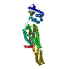
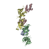
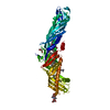
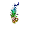
| Title | Crystal structure of the disulphide linked knotted homodimer of Psu | ||||||
 Components Components | Polarity suppression protein | ||||||
 Keywords Keywords | TRANSCRIPTION REGULATOR / All alpha protein / transcription termination inhibitor / Rho binding / capsid decoration protein of bacteriophage P4 / Transcription terminator Rho helicase | ||||||
| Function / homology | Phage polarity suppression protein monomer / symbiont-mediated activation of host transcription / Bacteriophage P4, Psu, polarity suppression protein / Phage polarity suppression protein (Psu) / Methane Monooxygenase Hydroxylase; Chain G, domain 1 / Up-down Bundle / Mainly Alpha / Polarity suppression protein Function and homology information Function and homology information | ||||||
| Biological species |  Enterobacteria phage P4 (virus) Enterobacteria phage P4 (virus) | ||||||
| Method |  X-RAY DIFFRACTION / MOLECULAR REPLACEMENT / Resolution: 3 Å X-RAY DIFFRACTION / MOLECULAR REPLACEMENT / Resolution: 3 Å | ||||||
 Authors Authors | Banerjee, R. / Nath, S. / Sen, U. | ||||||
 Citation Citation | Journal: J.Biol.Chem. / Year: 2012 Title: The first structure of polarity suppression protein, Psu from enterobacteria phage P4, reveals a novel fold and a knotted dimer Authors: Banerjee, R. / Nath, S. / Ranjan, A. / Khamrui, S. / Pani, B. / Sen, R. / Sen, U. | ||||||
| History |
|
- Structure visualization
| Structure viewer | Molecule: MolmilJmol/JSmol |
|---|
- Downloads & links
-Download
| PDBx/mmCIF format | 4dvd.cif.gz | 47.9 KB | Display | PDBx/mmCIF format |
|---|---|---|---|---|
| PDB format | pdb4dvd.ent.gz | 34.7 KB | Display | PDB format |
| PDBx/mmJSON format | 4dvd.json.gz | Tree view | PDBx/mmJSON format | |
| Others |  Other downloads Other downloads |
-Validation report
| Arichive directory | https://data.pdbj.org/pub/pdb/validation_reports/dv/4dvdftp://data.pdbj.org/pub/pdb/validation_reports/dv/4dvd | HTTPS FTP |
|---|
-Related structure data
| Related structure data |  3rx6SC S: Starting model for refinement C: citing same article ( |
|---|---|
| Similar structure data |


-Links
 PDBj
PDBj- Assembly
| Deposited unit | 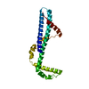
| ||||||||
|---|---|---|---|---|---|---|---|---|---|
| 1 | 
| ||||||||
| Unit cell |
|
-Components
| #1: Protein | Mass: 21362.973 Da / Num. of mol.: 1 / Mutation: C13S,C117S,T123C Source method: isolated from a genetically manipulated source Source: (gene. exp.) Enterobacteria phage P4 (virus) / Gene: psu / Plasmid: pET28a+ / Production host:  |
|---|---|
| Has protein modification | Y |
-Experimental details
-Experiment
| Experiment | Method: X-RAY DIFFRACTION / Number of used crystals: 1 |
|---|
- Sample preparation
| Crystal | Density Matthews: 4.11 Å3/Da / Density % sol: 70.04 % |
|---|---|
| Crystal grow | Temperature: 277 K / Method: vapor diffusion, hanging drop / pH: 7 Details: 7.5%(w/v) PEG 6000, 5%(v/v) glycerol, 0.1M HEPES, pH 7.0, VAPOR DIFFUSION, HANGING DROP, temperature 277K |
-Data collection
| Diffraction | Mean temperature: 100 K |
|---|---|
| Diffraction source | Source: ROTATING ANODE / Type: ENRAF-NONIUS FR591 / Wavelength: 1.5418 Å |
| Detector | Type: MAR scanner 345 mm plate / Detector: IMAGE PLATE / Date: Jan 15, 2012 / Details: Osmic maxflux |
| Radiation | Monochromator: Ni FILTER / Protocol: SINGLE WAVELENGTH / Monochromatic (M) / Laue (L): M / Scattering type: x-ray |
| Radiation wavelength | Wavelength: 1.5418 Å / Relative weight: 1 |
| Reflection | Resolution: 3→47.311 Å / Num. obs: 6846 / % possible obs: 94.1 % / Redundancy: 2.9 % / Biso Wilson estimate: 51.09 Å2 / Rmerge(I) obs: 0.115 / Net I/σ(I): 5.8 |
| Reflection shell | Resolution: 3→3.16 Å / % possible all: 93.7 |
- Processing
| Software |
| ||||||||||||||||||||||||
|---|---|---|---|---|---|---|---|---|---|---|---|---|---|---|---|---|---|---|---|---|---|---|---|---|---|
| Refinement | Method to determine structure: MOLECULAR REPLACEMENT Starting model: 3RX6 Resolution: 3→47.311 Å / Occupancy max: 1 / Occupancy min: 0.49 / FOM work R set: 0.7767 / SU ML: 0.61 / Isotropic thermal model: Isotropic / Cross valid method: THROUGHOUT / σ(F): 1.34 / Phase error: 28.36 / Stereochemistry target values: ML
| ||||||||||||||||||||||||
| Solvent computation | Shrinkage radii: 0.83 Å / VDW probe radii: 1.1 Å / Solvent model: FLAT BULK SOLVENT MODEL / Bsol: 21.695 Å2 / ksol: 0.334 e/Å3 | ||||||||||||||||||||||||
| Displacement parameters | Biso max: 136.92 Å2 / Biso mean: 58.1161 Å2 / Biso min: 21.27 Å2
| ||||||||||||||||||||||||
| Refinement step | Cycle: LAST / Resolution: 3→47.311 Å
| ||||||||||||||||||||||||
| Refine LS restraints |
| ||||||||||||||||||||||||
| LS refinement shell | Refine-ID: X-RAY DIFFRACTION / Total num. of bins used: 2
|
