 Movie
Movie Controller

[English] 日本語
 Yorodumi
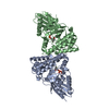
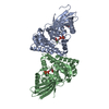
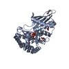
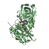
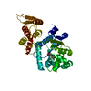
- PDB-4du7: Crystal structure of Staphylococcus epidermidis mevalonate diphos... -
+ Open data

- Basic information
| Entry | Database: PDB / ID: 4du7 | ||||||
|---|---|---|---|---|---|---|---|
| Title | Crystal structure of Staphylococcus epidermidis mevalonate diphosphate decarboxylase complexed with substrate mevalonate diphosphate | ||||||
 Components Components | Mevalonate diphosphate decarboxylase | ||||||
 Keywords Keywords | LYASE / GHMP Kinase Family | ||||||
| Function / homology |  Function and homology information Function and homology informationdiphosphomevalonate decarboxylase / diphosphomevalonate decarboxylase activity / isopentenyl diphosphate biosynthetic process, mevalonate pathway / ATP binding / cytosol Similarity search - Function | ||||||
| Biological species |   Staphylococcus epidermidis (bacteria) Staphylococcus epidermidis (bacteria) | ||||||
| Method |  X-RAY DIFFRACTION / SYNCHROTRON / MOLECULAR REPLACEMENT / Resolution: 2.201 Å X-RAY DIFFRACTION / SYNCHROTRON / MOLECULAR REPLACEMENT / Resolution: 2.201 Å | ||||||
 Authors Authors | Barta, M.L. / McWhorter, W.J. / Geisbrecht, B.V. | ||||||
 Citation Citation | Journal: Biochemistry / Year: 2012 Title: Structural basis for nucleotide binding and reaction catalysis in mevalonate diphosphate decarboxylase. Authors: Barta, M.L. / McWhorter, W.J. / Miziorko, H.M. / Geisbrecht, B.V. | ||||||
| History |
|
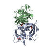
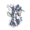
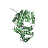
- Structure visualization
| Structure viewer | Molecule: MolmilJmol/JSmol |
|---|
- Downloads & links
-Download
| PDBx/mmCIF format | 4du7.cif.gz | 143 KB | Display | PDBx/mmCIF format |
|---|---|---|---|---|
| PDB format | pdb4du7.ent.gz | 112 KB | Display | PDB format |
| PDBx/mmJSON format | 4du7.json.gz | Tree view | PDBx/mmJSON format | |
| Others |  Other downloads Other downloads |
-Validation report
| Arichive directory | https://data.pdbj.org/pub/pdb/validation_reports/du/4du7ftp://data.pdbj.org/pub/pdb/validation_reports/du/4du7 | HTTPS FTP |
|---|
-Related structure data
| Related structure data | 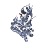 4dptC 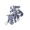 4dpuC 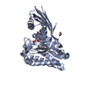 4dpwC 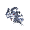 4dpxC 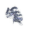 4dpyC  4du8C 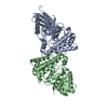 3qt5S S: Starting model for refinement C: citing same article ( |
|---|---|
| Similar structure data |


-Links
 PDBj
PDBj- Assembly
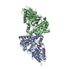
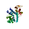
| Deposited unit | 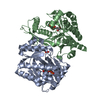
| ||||||||
|---|---|---|---|---|---|---|---|---|---|
| 1 | 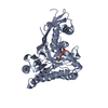
| ||||||||
| 2 | 
| ||||||||
| Unit cell |
|
-Components
| #1: Protein | Mass: 36870.395 Da / Num. of mol.: 2 Source method: isolated from a genetically manipulated source Source: (gene. exp.) Staphylococcus epidermidis (bacteria) / Gene: mvaD / Plasmid: pT7HMT / Production host: References: UniProt: Q9FD73, diphosphomevalonate decarboxylase #2: Chemical | 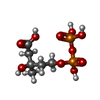  Mass: 308.117 Da / Num. of mol.: 2 / Source method: obtained synthetically / Formula: C6H14O10P2 Mass: 308.117 Da / Num. of mol.: 2 / Source method: obtained synthetically / Formula: C6H14O10P2#3: Chemical | ChemComp-FMT / |   Mass: 46.025 Da / Num. of mol.: 1 / Source method: obtained synthetically / Formula: CH2O2 Mass: 46.025 Da / Num. of mol.: 1 / Source method: obtained synthetically / Formula: CH2O2#4: Water | ChemComp-HOH / |  Mass: 18.015 Da / Num. of mol.: 272 / Source method: isolated from a natural source / Formula: H2O Mass: 18.015 Da / Num. of mol.: 272 / Source method: isolated from a natural source / Formula: H2O |
|---|
-Experimental details
-Experiment
| Experiment | Method: X-RAY DIFFRACTION / Number of used crystals: 1 |
|---|
- Sample preparation
| Crystal | Density Matthews: 2.2 Å3/Da / Density % sol: 44.03 % |
|---|---|
| Crystal grow | Temperature: 298 K / Method: vapor diffusion, hanging drop / pH: 7 Details: 0.25 M sodium formate, 16% w/v PEG3350, pH 7.0, VAPOR DIFFUSION, HANGING DROP, temperature 298K |
-Data collection
| Diffraction | Mean temperature: 93 K |
|---|---|
| Diffraction source | Source: SYNCHROTRON / Site: APS  / Beamline: 23-BM-B / Wavelength: 1 Å / Beamline: 23-BM-B / Wavelength: 1 Å |
| Detector | Type: MARMOSAIC 225 mm CCD / Detector: CCD / Date: Dec 12, 2011 / Details: mirror |
| Radiation | Monochromator: Si(111) / Protocol: SINGLE WAVELENGTH / Monochromatic (M) / Laue (L): M / Scattering type: x-ray |
| Radiation wavelength | Wavelength: 1 Å / Relative weight: 1 |
| Reflection | Resolution: 2.2→50 Å / Num. all: 31892 / Num. obs: 30297 / % possible obs: 95.7 % / Observed criterion σ(F): 0 / Observed criterion σ(I): 2 / Redundancy: 9.2 % / Biso Wilson estimate: 30.71 Å2 / Rmerge(I) obs: 0.117 / Net I/σ(I): 19.8 |
| Reflection shell | Resolution: 2.2→2.2716 Å / Rmerge(I) obs: 0.557 / Mean I/σ(I) obs: 3.04 / % possible all: 88 |
- Processing
| Software |
| ||||||||||||||||||||||||||||||||||||||||||||||||||||||||||||||||||||||||||||||||||||
|---|---|---|---|---|---|---|---|---|---|---|---|---|---|---|---|---|---|---|---|---|---|---|---|---|---|---|---|---|---|---|---|---|---|---|---|---|---|---|---|---|---|---|---|---|---|---|---|---|---|---|---|---|---|---|---|---|---|---|---|---|---|---|---|---|---|---|---|---|---|---|---|---|---|---|---|---|---|---|---|---|---|---|---|---|---|
| Refinement | Method to determine structure: MOLECULAR REPLACEMENT Starting model: PDB ENTRY 3QT5 Resolution: 2.201→31.261 Å / Occupancy max: 1 / Occupancy min: 0.69 / FOM work R set: 0.8095 / SU ML: 0.28 / σ(F): 0 / Phase error: 25.71 / Stereochemistry target values: ML
| ||||||||||||||||||||||||||||||||||||||||||||||||||||||||||||||||||||||||||||||||||||
| Solvent computation | Shrinkage radii: 0.95 Å / VDW probe radii: 1.2 Å / Solvent model: FLAT BULK SOLVENT MODEL / Bsol: 33.724 Å2 / ksol: 0.347 e/Å3 | ||||||||||||||||||||||||||||||||||||||||||||||||||||||||||||||||||||||||||||||||||||
| Displacement parameters | Biso max: 99.52 Å2 / Biso mean: 34.9367 Å2 / Biso min: 17.52 Å2
| ||||||||||||||||||||||||||||||||||||||||||||||||||||||||||||||||||||||||||||||||||||
| Refinement step | Cycle: LAST / Resolution: 2.201→31.261 Å
| ||||||||||||||||||||||||||||||||||||||||||||||||||||||||||||||||||||||||||||||||||||
| Refine LS restraints |
| ||||||||||||||||||||||||||||||||||||||||||||||||||||||||||||||||||||||||||||||||||||
| LS refinement shell | Refine-ID: X-RAY DIFFRACTION / Total num. of bins used: 11
|
