 Movie
Movie Controller

[English] 日本語
 Yorodumi
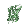
- PDB-2qdl: Crystal structure of scaffolding protein TtCheW from Thermoanaero... -
+ Open data

- Basic information
| Entry | Database: PDB / ID: 2qdl | ||||||
|---|---|---|---|---|---|---|---|
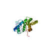
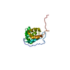
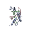
| Title | Crystal structure of scaffolding protein TtCheW from Thermoanaerobacter tengcongensis | ||||||
 Components Components | Chemotaxis signal transduction protein | ||||||
 Keywords Keywords | SIGNALING PROTEIN / beta-barrel | ||||||
| Function / homology |  Function and homology information Function and homology information | ||||||
| Biological species |  Thermoanaerobacter tengcongensis (bacteria) Thermoanaerobacter tengcongensis (bacteria) | ||||||
| Method |  X-RAY DIFFRACTION / MOLECULAR REPLACEMENT / Resolution: 2.2 Å X-RAY DIFFRACTION / MOLECULAR REPLACEMENT / Resolution: 2.2 Å | ||||||
 Authors Authors | Yao, W. / Shi, L. / Liang, D.C. | ||||||
 Citation Citation | Journal: Biochem.Biophys.Res.Commun. / Year: 2007 Title: Crystal structure of scaffolding protein CheW from thermoanaerobacter tengcongensis. Authors: Yao, W. / Shi, L. / Liang, D.C. | ||||||
| History |
|
- Structure visualization
| Structure viewer | Molecule: MolmilJmol/JSmol |
|---|
- Downloads & links
-Download
| PDBx/mmCIF format | 2qdl.cif.gz | 72 KB | Display | PDBx/mmCIF format |
|---|---|---|---|---|
| PDB format | pdb2qdl.ent.gz | 53.9 KB | Display | PDB format |
| PDBx/mmJSON format | 2qdl.json.gz | Tree view | PDBx/mmJSON format | |
| Others |  Other downloads Other downloads |
-Validation report
| Arichive directory | https://data.pdbj.org/pub/pdb/validation_reports/qd/2qdlftp://data.pdbj.org/pub/pdb/validation_reports/qd/2qdl | HTTPS FTP |
|---|
-Related structure data
| Related structure data |  2ch4S S: Starting model for refinement |
|---|---|
| Similar structure data |


-Links
 PDBj
PDBj- Assembly
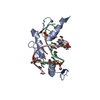
| Deposited unit | 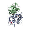
| ||||||||
|---|---|---|---|---|---|---|---|---|---|
| 1 | 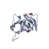
| ||||||||
| 2 | 
| ||||||||
| Unit cell |
| ||||||||
| Details | The biological assembly is a monomer. |
-Components
| #1: Protein | Mass: 18554.744 Da / Num. of mol.: 2 Source method: isolated from a genetically manipulated source Source: (gene. exp.) Thermoanaerobacter tengcongensis (bacteria)Strain: MB4 / Plasmid: pETM10 / Species (production host): Escherichia coli / Production host: #2: Chemical |   Mass: 92.094 Da / Num. of mol.: 3 / Source method: obtained synthetically / Formula: C3H8O3 Mass: 92.094 Da / Num. of mol.: 3 / Source method: obtained synthetically / Formula: C3H8O3#3: Water | ChemComp-HOH / |  Mass: 18.015 Da / Num. of mol.: 103 / Source method: isolated from a natural source / Formula: H2O Mass: 18.015 Da / Num. of mol.: 103 / Source method: isolated from a natural source / Formula: H2O |
|---|
-Experimental details
-Experiment
| Experiment | Method: X-RAY DIFFRACTION |
|---|
- Sample preparation
| Crystal | Density Matthews: 2.14 Å3/Da / Density % sol: 42.48 % |
|---|---|
| Crystal grow | Temperature: 293 K / Method: vapor diffusion, hanging drop / pH: 5.4 Details: 0.1M acetate buffer (pH 5.4), 0.2M Sodium Nitrate, 10% (w/v) PEG 3350, VAPOR DIFFUSION, HANGING DROP, temperature 293.0K |
-Data collection
| Diffraction | Mean temperature: 100 K |
|---|---|
| Diffraction source | Source: ROTATING ANODE / Type: RIGAKU / Wavelength: 1.5418 Å |
| Detector | Type: RIGAKU RAXIS IV / Detector: IMAGE PLATE / Date: Oct 17, 2006 |
| Radiation | Protocol: SINGLE WAVELENGTH / Monochromatic (M) / Laue (L): M / Scattering type: x-ray |
| Radiation wavelength | Wavelength: 1.5418 Å / Relative weight: 1 |
| Reflection | Resolution: 2.2→19.8 Å / Num. obs: 14589 / % possible obs: 96 % / Redundancy: 7.3 % / Rmerge(I) obs: 0.078 |
| Reflection shell | Resolution: 2.2→2.25 Å / Rmerge(I) obs: 0.286 / % possible all: 93.6 |
- Processing
| Software |
| ||||||||||||||||||||||||||||||||||||||||||||||||||||||||||||||||||||||||||||||||||||||||||
|---|---|---|---|---|---|---|---|---|---|---|---|---|---|---|---|---|---|---|---|---|---|---|---|---|---|---|---|---|---|---|---|---|---|---|---|---|---|---|---|---|---|---|---|---|---|---|---|---|---|---|---|---|---|---|---|---|---|---|---|---|---|---|---|---|---|---|---|---|---|---|---|---|---|---|---|---|---|---|---|---|---|---|---|---|---|---|---|---|---|---|---|
| Refinement | Method to determine structure: MOLECULAR REPLACEMENT Starting model: PDB entry 2CH4 Resolution: 2.2→19.76 Å / Cor.coef. Fo:Fc: 0.941 / Cor.coef. Fo:Fc free: 0.92 / SU B: 5.504 / SU ML: 0.143 / Cross valid method: THROUGHOUT / ESU R: 0.319 / ESU R Free: 0.217 / Stereochemistry target values: MAXIMUM LIKELIHOOD / Details: HYDROGENS HAVE BEEN ADDED IN THE RIDING POSITIONS
| ||||||||||||||||||||||||||||||||||||||||||||||||||||||||||||||||||||||||||||||||||||||||||
| Solvent computation | Ion probe radii: 0.8 Å / Shrinkage radii: 0.8 Å / VDW probe radii: 1.2 Å / Solvent model: MASK | ||||||||||||||||||||||||||||||||||||||||||||||||||||||||||||||||||||||||||||||||||||||||||
| Displacement parameters | Biso mean: 21.877 Å2
| ||||||||||||||||||||||||||||||||||||||||||||||||||||||||||||||||||||||||||||||||||||||||||
| Refinement step | Cycle: LAST / Resolution: 2.2→19.76 Å
| ||||||||||||||||||||||||||||||||||||||||||||||||||||||||||||||||||||||||||||||||||||||||||
| Refine LS restraints |
| ||||||||||||||||||||||||||||||||||||||||||||||||||||||||||||||||||||||||||||||||||||||||||
| LS refinement shell | Resolution: 2.2→2.256 Å / Total num. of bins used: 20
|
