Mass: 18.015 Da / Num. of mol.: 438 / Source method: isolated from a natural source / Formula: H2O
Has protein modification
Y
Sequence details
THE CONSTRUCT (RESIDUES 27-380) WAS EXPRESSED WITH AN N- TERMINAL PURIFICATION TAG ...THE CONSTRUCT (RESIDUES 27-380) WAS EXPRESSED WITH AN N- TERMINAL PURIFICATION TAG MGSDKIHHHHHHENLYFQG. THE TAG WAS REMOVED WITH TEV PROTEASE LEAVING ONLY A GLYCINE (0) FOLLOWED BY THE TARGET SEQUENCE.
-
Experimental details
-
Experiment
Experiment
Method: X-RAY DIFFRACTION / Number of used crystals: 1
-
Sample preparation
Crystal
Density Matthews: 2.53 Å3/Da / Density % sol: 51.34 %
Crystal grow
Temperature: 293 K / Method: vapor diffusion, sitting drop / pH: 7 Details: 0.2M sodium chloride, 1.0M sodium citrate, 0.1M TRIS pH 7.0, NANODROP, VAPOR DIFFUSION, SITTING DROP, temperature 293K
Monochromator: double crystal Si(111) / Protocol: SINGLE WAVELENGTH / Monochromatic (M) / Laue (L): M / Scattering type: x-ray
Radiation wavelength
Wavelength: 0.97916 Å / Relative weight: 1
Reflection
Resolution: 1.5→29.183 Å / Num. all: 65989 / Num. obs: 65989 / % possible obs: 100 % / Redundancy: 9 % / Rsym value: 0.088 / Net I/σ(I): 11.7
Reflection shell
Diffraction-ID: 1
Resolution (Å)
Redundancy (%)
Rmerge(I) obs
Mean I/σ(I) obs
Num. measured all
Num. unique all
Rsym value
% possible all
1.5-1.54
8.9
0.848
0.9
43006
4810
0.848
100
1.54-1.58
9
0.719
1.1
41695
4646
0.719
100
1.58-1.63
9
0.548
1.4
40998
4566
0.548
100
1.63-1.68
9
0.439
1.8
39732
4409
0.439
100
1.68-1.73
9
0.357
2.2
38642
4286
0.357
100
1.73-1.79
9
0.28
2.8
37761
4183
0.28
100
1.79-1.86
9
0.22
3.5
36329
4021
0.22
100
1.86-1.94
9.1
0.169
4.4
35034
3865
0.169
100
1.94-2.02
9.1
0.141
5.2
33697
3721
0.141
100
2.02-2.12
9
0.126
5.5
32323
3579
0.126
100
2.12-2.24
9
0.118
5.7
30751
3406
0.118
100
2.24-2.37
9
0.118
5.7
29245
3233
0.118
100
2.37-2.54
9
0.107
6.1
27578
3051
0.107
100
2.54-2.74
9
0.092
7.2
25592
2834
0.092
100
2.74-3
9
0.077
8.6
23855
2650
0.077
100
3-3.35
8.9
0.066
9.9
21458
2404
0.066
100
3.35-3.87
8.9
0.058
11.2
18979
2144
0.058
100
3.87-4.74
8.7
0.053
12.5
16006
1845
0.053
100
4.74-6.71
8.4
0.06
10.9
12357
1473
0.06
99.9
6.71-29.183
7.4
0.065
9.9
6378
863
0.065
97.4
-
Phasing
Phasing
Method: SAD
-
Processing
Software
Name
Version
Classification
NB
MolProbity
3beta29
modelbuilding
PDB_EXTRACT
3.1
dataextraction
SHELX
phasing
SHARP
phasing
SCALA
3.3.20
datascaling
PHENIX
1.7.3
refinement
MOSFLM
datareduction
SHELXD
phasing
Refinement
Method to determine structure: SAD / Resolution: 1.5→29.183 Å / Occupancy max: 1 / Occupancy min: 0.25 / SU ML: 0.18 / σ(F): 1.34 / Phase error: 15.3 / Stereochemistry target values: MLHL Details: 1. HYDROGENS HAVE BEEN ADDED IN THE RIDING POSITIONS. 2. A MET-INHIBITION PROTOCOL WAS USED FOR SELENOMETHIONINE INCORPORATION DURING PROTEIN EXPRESSION. THE OCCUPANCY OF THE SE ATOMS IN THE ...Details: 1. HYDROGENS HAVE BEEN ADDED IN THE RIDING POSITIONS. 2. A MET-INHIBITION PROTOCOL WAS USED FOR SELENOMETHIONINE INCORPORATION DURING PROTEIN EXPRESSION. THE OCCUPANCY OF THE SE ATOMS IN THE MSE RESIDUES WAS REDUCED TO 0.75 FOR THE REDUCED SCATTERING POWER DUE TO PARTIAL S-MET INCORPORATION. 3. ATOM RECORD CONTAINS SUM OF TLS AND RESIDUAL B FACTORS. ANISOU RECORD CONTAINS SUM OF TLS AND RESIDUAL U FACTORS. 4. WATERS WERE EXCLUDED FROM AUTOMATIC TLS ASSIGNMENT.
Rfactor
Num. reflection
% reflection
Rfree
0.1764
3343
5.07 %
Rwork
0.1558
-
-
obs
0.1569
65902
99.96 %
Solvent computation
Shrinkage radii: 0.6 Å / VDW probe radii: 0.9 Å / Solvent model: FLAT BULK SOLVENT MODEL / Bsol: 44.188 Å2 / ksol: 0.41 e/Å3
In the structure databanks used in Yorodumi, some data are registered as the other names, "COVID-19 virus" and "2019-nCoV". Here are the details of the virus and the list of structure data.
Jan 31, 2019. EMDB accession codes are about to change! (news from PDBe EMDB page)
EMDB accession codes are about to change! (news from PDBe EMDB page)
The allocation of 4 digits for EMDB accession codes will soon come to an end. Whilst these codes will remain in use, new EMDB accession codes will include an additional digit and will expand incrementally as the available range of codes is exhausted. The current 4-digit format prefixed with “EMD-” (i.e. EMD-XXXX) will advance to a 5-digit format (i.e. EMD-XXXXX), and so on. It is currently estimated that the 4-digit codes will be depleted around Spring 2019, at which point the 5-digit format will come into force.
The EM Navigator/Yorodumi systems omit the EMD- prefix.
Related info.:Q: What is EMD? / ID/Accession-code notation in Yorodumi/EM Navigator
Yorodumi is a browser for structure data from EMDB, PDB, SASBDB, etc.
This page is also the successor to EM Navigator detail page, and also detail information page/front-end page for Omokage search.
The word "yorodu" (or yorozu) is an old Japanese word meaning "ten thousand". "mi" (miru) is to see.
Related info.:EMDB / PDB / SASBDB / Comparison of 3 databanks / Yorodumi Search / Aug 31, 2016. New EM Navigator & Yorodumi / Yorodumi Papers / Jmol/JSmol / Function and homology information / Changes in new EM Navigator and Yorodumi
 Movie
Movie Controller

 Yorodumi
 Open data
 Basic information
 Components
 Keywords
 Function and homology information
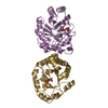
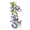
 Bacteroides ovatus (bacteria)
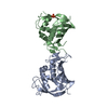
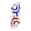
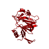
 X-RAY DIFFRACTION /
  Authors
 Citation
 Structure visualization
 Downloads & links
 Other downloads
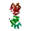


 PDBj
PDBj Assembly
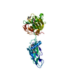
 Mass: 18.015 Da / Num. of mol.: 438 / Source method: isolated from a natural source / Formula: H2O
 Sample preparation
 / Beamline: BL14-1 / Wavelength: 0.97916
  Processing