- PDB-4dnn: Crystal structure of the Quaking Qua1 homodimerization domain -
+
データを開く
IDまたはキーワード:
読み込み中...
-
基本情報
登録情報
データベース: PDB / ID: 4dnn
タイトル
Crystal structure of the Quaking Qua1 homodimerization domain
要素
Protein quaking
キーワード
SPLICING / Helix-turn-helix / HYDROPHOBIC HOMODIMER INTERFACE / PERPENDICULAR STACKING OF Protomers / DEVELOPMENTAL PROTEIN / RNA-binding / TRANSLATION REGULATION
機能・相同性
機能・相同性情報
internal N(7)-methylguanine-containing RNA reader activity / positive regulation of 3'-UTR-mediated mRNA stabilization / negative regulation of miRNA catabolic process / myofibroblast contraction / negative regulation of 3'-UTR-mediated mRNA stabilization / spliceosome-depend formation of circular RNA / regulation of astrocyte differentiation / negative regulation of macrophage differentiation / axon ensheathment / vascular associated smooth muscle cell differentiation ...internal N(7)-methylguanine-containing RNA reader activity / positive regulation of 3'-UTR-mediated mRNA stabilization / negative regulation of miRNA catabolic process / myofibroblast contraction / negative regulation of 3'-UTR-mediated mRNA stabilization / spliceosome-depend formation of circular RNA / regulation of astrocyte differentiation / negative regulation of macrophage differentiation / axon ensheathment / vascular associated smooth muscle cell differentiation / 3'-UTR-mediated mRNA destabilization / regulation of epithelial to mesenchymal transition / positive regulation of myelination / intracellular mRNA localization / microglia differentiation / regulation of mRNA splicing, via spliceosome / negative regulation of cold-induced thermogenesis / long-chain fatty acid biosynthetic process / miRNA binding / mRNA stabilization / negative regulation of type I interferon production / positive regulation of oligodendrocyte differentiation / spermatid development / mRNA transport / vasculogenesis / myelination / negative regulation of angiogenesis / mRNA 3'-UTR binding / positive regulation of cholesterol biosynthetic process / SH3 domain binding / cytoplasmic stress granule / transcription coactivator activity / negative regulation of translation / mRNA binding / positive regulation of gene expression / synapse / DNA binding / RNA binding / nucleus / cytoplasm / cytosol 類似検索 - 分子機能
Single alpha-helices involved in coiled-coils or other helix-helix interfaces - #4010 / Protein quaking, putative nuclear localisation signal / Putative nuclear localisation signal of quaking / STAR protein, homodimerisation region / Homodimerisation region of STAR domain protein / : / KHDC4/BBP-like, KH-domain type I / KH domain-containing BBP-like / K Homology domain, type 1 superfamily / Single alpha-helices involved in coiled-coils or other helix-helix interfaces ...Single alpha-helices involved in coiled-coils or other helix-helix interfaces - #4010 / Protein quaking, putative nuclear localisation signal / Putative nuclear localisation signal of quaking / STAR protein, homodimerisation region / Homodimerisation region of STAR domain protein / : / KHDC4/BBP-like, KH-domain type I / KH domain-containing BBP-like / K Homology domain, type 1 superfamily / Single alpha-helices involved in coiled-coils or other helix-helix interfaces / K Homology domain / K homology RNA-binding domain / Up-down Bundle / Mainly Alpha 類似検索 - ドメイン・相同性
KH domain-containing RNA-binding protein QKI 類似検索 - 構成要素
 ムービー
ムービー コントローラー


 データを開く
 基本情報
基本情報 要素
要素 キーワード
キーワード 機能・相同性情報
 X線回折 /
  データ登録者
 引用
引用 構造の表示
構造の表示 ダウンロードとリンク
 その他のダウンロード
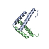


 PDBj
PDBj
 集合体
集合体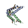


 分子量: 40.078 Da / 分子数: 1 / 由来タイプ: 合成 / 式: Ca
 分子量: 18.015 Da / 分子数: 44 / 由来タイプ: 天然 / 式: H2O
 試料調製
試料調製 / ビームライン: BL11-1 / 波長: 0.9791358, 0.9184018, 0.979569
 解析
解析