- PDB-4dnn: Crystal structure of the Quaking Qua1 homodimerization domain -
+
Open data
ID or keywords:
Loading...
-
Basic information
Entry
Database: PDB / ID: 4dnn
Title
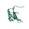
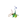
Crystal structure of the Quaking Qua1 homodimerization domain
Components
Protein quaking
Keywords
SPLICING / Helix-turn-helix / HYDROPHOBIC HOMODIMER INTERFACE / PERPENDICULAR STACKING OF Protomers / DEVELOPMENTAL PROTEIN / RNA-binding / TRANSLATION REGULATION
Function / homology
Function and homology information
internal N(7)-methylguanine-containing RNA reader activity / positive regulation of 3'-UTR-mediated mRNA stabilization / negative regulation of miRNA catabolic process / myofibroblast contraction / negative regulation of 3'-UTR-mediated mRNA stabilization / spliceosome-depend formation of circular RNA / regulation of astrocyte differentiation / negative regulation of macrophage differentiation / axon ensheathment / vascular associated smooth muscle cell differentiation ...internal N(7)-methylguanine-containing RNA reader activity / positive regulation of 3'-UTR-mediated mRNA stabilization / negative regulation of miRNA catabolic process / myofibroblast contraction / negative regulation of 3'-UTR-mediated mRNA stabilization / spliceosome-depend formation of circular RNA / regulation of astrocyte differentiation / negative regulation of macrophage differentiation / axon ensheathment / vascular associated smooth muscle cell differentiation / 3'-UTR-mediated mRNA destabilization / regulation of epithelial to mesenchymal transition / positive regulation of myelination / intracellular mRNA localization / microglia differentiation / regulation of mRNA splicing, via spliceosome / negative regulation of cold-induced thermogenesis / long-chain fatty acid biosynthetic process / miRNA binding / mRNA stabilization / negative regulation of type I interferon production / positive regulation of oligodendrocyte differentiation / spermatid development / vasculogenesis / mRNA transport / myelination / negative regulation of angiogenesis / mRNA 3'-UTR binding / positive regulation of cholesterol biosynthetic process / SH3 domain binding / cytoplasmic stress granule / transcription coactivator activity / negative regulation of translation / mRNA binding / positive regulation of gene expression / synapse / DNA binding / RNA binding / nucleus / cytoplasm / cytosol Similarity search - Function
Single alpha-helices involved in coiled-coils or other helix-helix interfaces - #4010 / Protein quaking, putative nuclear localisation signal / Putative nuclear localisation signal of quaking / STAR protein, homodimerisation region / Homodimerisation region of STAR domain protein / : / KHDC4/BBP-like, KH-domain type I / KH domain-containing BBP-like / K Homology domain, type 1 superfamily / Single alpha-helices involved in coiled-coils or other helix-helix interfaces ...Single alpha-helices involved in coiled-coils or other helix-helix interfaces - #4010 / Protein quaking, putative nuclear localisation signal / Putative nuclear localisation signal of quaking / STAR protein, homodimerisation region / Homodimerisation region of STAR domain protein / : / KHDC4/BBP-like, KH-domain type I / KH domain-containing BBP-like / K Homology domain, type 1 superfamily / Single alpha-helices involved in coiled-coils or other helix-helix interfaces / K Homology domain / K homology RNA-binding domain / Up-down Bundle / Mainly Alpha Similarity search - Domain/homology
In the structure databanks used in Yorodumi, some data are registered as the other names, "COVID-19 virus" and "2019-nCoV". Here are the details of the virus and the list of structure data.
Jan 31, 2019. EMDB accession codes are about to change! (news from PDBe EMDB page)
EMDB accession codes are about to change! (news from PDBe EMDB page)
The allocation of 4 digits for EMDB accession codes will soon come to an end. Whilst these codes will remain in use, new EMDB accession codes will include an additional digit and will expand incrementally as the available range of codes is exhausted. The current 4-digit format prefixed with “EMD-” (i.e. EMD-XXXX) will advance to a 5-digit format (i.e. EMD-XXXXX), and so on. It is currently estimated that the 4-digit codes will be depleted around Spring 2019, at which point the 5-digit format will come into force.
The EM Navigator/Yorodumi systems omit the EMD- prefix.
Related info.:Q: What is EMD? / ID/Accession-code notation in Yorodumi/EM Navigator
Yorodumi is a browser for structure data from EMDB, PDB, SASBDB, etc.
This page is also the successor to EM Navigator detail page, and also detail information page/front-end page for Omokage search.
The word "yorodu" (or yorozu) is an old Japanese word meaning "ten thousand". "mi" (miru) is to see.
Related info.:EMDB / PDB / SASBDB / Comparison of 3 databanks / Yorodumi Search / Aug 31, 2016. New EM Navigator & Yorodumi / Yorodumi Papers / Jmol/JSmol / Function and homology information / Changes in new EM Navigator and Yorodumi
 Movie
Movie Controller


 Open data
 Basic information
 Components
 Keywords
 Function and homology information
 X-RAY DIFFRACTION /
  Authors
 Citation
 Structure visualization
 Downloads & links
 Other downloads
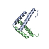


 PDBj
PDBj
 Assembly
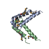


 Mass: 40.078 Da / Num. of mol.: 1 / Source method: obtained synthetically / Formula: Ca
 Mass: 18.015 Da / Num. of mol.: 44 / Source method: isolated from a natural source / Formula: H2O
 Sample preparation
 / Beamline: BL11-1 / Wavelength: 0.9791358, 0.9184018, 0.979569
 Processing