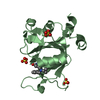
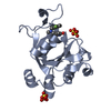
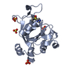
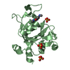
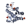
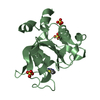
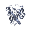
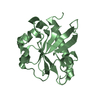
positive regulation of transcription elongation by RNA polymerase I / RNA polymerase II C-terminal domain phosphoserine binding / regulation of transcription-coupled nucleotide-excision repair / Cdc73/Paf1 complex / negative regulation of DNA recombination / mRNA 3'-end processing / RNA polymerase II complex binding / transcription elongation by RNA polymerase I / positive regulation of transcription elongation by RNA polymerase II / transcription elongation by RNA polymerase II ...positive regulation of transcription elongation by RNA polymerase I / RNA polymerase II C-terminal domain phosphoserine binding / regulation of transcription-coupled nucleotide-excision repair / Cdc73/Paf1 complex / negative regulation of DNA recombination / mRNA 3'-end processing / RNA polymerase II complex binding / transcription elongation by RNA polymerase I / positive regulation of transcription elongation by RNA polymerase II / transcription elongation by RNA polymerase II / euchromatin / chromatin binding / nucleus Similarity search - Function
RNA polymerase II accessory factor, Cdc73 C-terminal domain / Cdc73/Parafibromin / Cell division control protein 73, C-terminal / Cell division control protein 73, C-terminal domain superfamily / RNA pol II accessory factor, Cdc73 family, C-terminal / Rossmann fold / 3-Layer(aba) Sandwich / Alpha Beta Similarity search - Domain/homology
Method to determine structure: SAD / Resolution: 2.19→50 Å / Cor.coef. Fo:Fc: 0.932 / Cor.coef. Fo:Fc free: 0.909 / SU B: 11.692 / SU ML: 0.135 / Cross valid method: THROUGHOUT / ESU R: 0.223 / ESU R Free: 0.199 / Stereochemistry target values: MAXIMUM LIKELIHOOD / Details: HYDROGENS HAVE BEEN ADDED IN THE RIDING POSITIONS
Rfactor
Num. reflection
% reflection
Selection details
Rfree
0.26505
1324
5.1 %
RANDOM
Rwork
0.22056
-
-
-
obs
0.22275
24765
99.64 %
-
all
-
26095
-
-
Solvent computation
Ion probe radii: 0.8 Å / Shrinkage radii: 0.8 Å / VDW probe radii: 1.4 Å / Solvent model: MASK
Displacement parameters
Biso mean: 32.347 Å2
Baniso -1
Baniso -2
Baniso -3
1-
1.13 Å2
0 Å2
0 Å2
2-
-
0.9 Å2
0 Å2
3-
-
-
-2.03 Å2
Refinement step
Cycle: LAST / Resolution: 2.19→50 Å
Protein
Nucleic acid
Ligand
Solvent
Total
Num. atoms
2588
0
0
187
2775
Refine LS restraints
Refine-ID
Type
Dev ideal
Dev ideal target
Number
X-RAY DIFFRACTION
r_bond_refined_d
0.008
0.022
2678
X-RAY DIFFRACTION
r_bond_other_d
X-RAY DIFFRACTION
r_angle_refined_deg
1.061
1.921
3649
X-RAY DIFFRACTION
r_angle_other_deg
X-RAY DIFFRACTION
r_dihedral_angle_1_deg
5.286
5
316
X-RAY DIFFRACTION
r_dihedral_angle_2_deg
39.818
23.111
135
X-RAY DIFFRACTION
r_dihedral_angle_3_deg
14.948
15
421
X-RAY DIFFRACTION
r_dihedral_angle_4_deg
21.865
15
20
X-RAY DIFFRACTION
r_chiral_restr
0.078
0.2
389
X-RAY DIFFRACTION
r_gen_planes_refined
0.004
0.021
2090
X-RAY DIFFRACTION
r_gen_planes_other
X-RAY DIFFRACTION
r_nbd_refined
X-RAY DIFFRACTION
r_nbd_other
X-RAY DIFFRACTION
r_nbtor_refined
X-RAY DIFFRACTION
r_nbtor_other
X-RAY DIFFRACTION
r_xyhbond_nbd_refined
X-RAY DIFFRACTION
r_xyhbond_nbd_other
X-RAY DIFFRACTION
r_metal_ion_refined
X-RAY DIFFRACTION
r_metal_ion_other
X-RAY DIFFRACTION
r_symmetry_vdw_refined
X-RAY DIFFRACTION
r_symmetry_vdw_other
X-RAY DIFFRACTION
r_symmetry_hbond_refined
X-RAY DIFFRACTION
r_symmetry_hbond_other
X-RAY DIFFRACTION
r_symmetry_metal_ion_refined
X-RAY DIFFRACTION
r_symmetry_metal_ion_other
X-RAY DIFFRACTION
r_mcbond_it
0.419
1.5
1590
X-RAY DIFFRACTION
r_mcbond_other
X-RAY DIFFRACTION
r_mcangle_it
0.8
2
2588
X-RAY DIFFRACTION
r_scbond_it
1.169
3
1088
X-RAY DIFFRACTION
r_scangle_it
1.915
4.5
1061
X-RAY DIFFRACTION
r_rigid_bond_restr
X-RAY DIFFRACTION
r_sphericity_free
X-RAY DIFFRACTION
r_sphericity_bonded
LS refinement shell
Resolution: 2.193→2.25 Å / Total num. of bins used: 20
Rfactor
Num. reflection
% reflection
Rfree
0.348
90
-
Rwork
0.288
1737
-
obs
-
-
95.8 %
Refinement TLS params.
Method: refined / Refine-ID: X-RAY DIFFRACTION
ID
L11 (°2)
L12 (°2)
L13 (°2)
L22 (°2)
L23 (°2)
L33 (°2)
S11 (Å °)
S12 (Å °)
S13 (Å °)
S21 (Å °)
S22 (Å °)
S23 (Å °)
S31 (Å °)
S32 (Å °)
S33 (Å °)
T11 (Å2)
T12 (Å2)
T13 (Å2)
T22 (Å2)
T23 (Å2)
T33 (Å2)
Origin x (Å)
Origin y (Å)
Origin z (Å)
1
2.289
0.128
1.2241
2.213
1.1002
4.0319
0.1693
0.0436
-0.0117
-0.1251
-0.0635
-0.0757
0.1902
0.1034
-0.1058
0.0701
-0.0123
0.0315
0.0658
0.0155
0.0593
10.9804
12.0625
38.4486
2
2.0687
-0.5487
-0.9008
1.1421
-0.1553
5.3731
0.1555
-0.0366
0.2095
-0.0668
0.0165
0.1005
-0.4966
-0.0397
-0.172
0.0973
0.0393
0.0088
0.0629
0.0427
0.125
-16.8035
21.6895
41.6425
3
1.3591
-0.0349
0.1403
0.4507
-0.3506
0.4443
0.0462
0.0288
0.1046
-0.0331
0.0098
0.0407
-0.0629
-0.021
-0.056
0.1163
0.005
0.0138
0.0903
0.0184
0.0863
-3.166
17.1683
40.6469
Refinement TLS group
ID
Refine-ID
Refine TLS-ID
Auth asym-ID
Auth seq-ID
1
X-RAY DIFFRACTION
1
A
2 - 160
2
X-RAY DIFFRACTION
2
B
4 - 160
3
X-RAY DIFFRACTION
3
A
201 - 283
4
X-RAY DIFFRACTION
3
B
201 - 304
+
About Yorodumi
-
News
-
Feb 9, 2022. New format data for meta-information of EMDB entries
New format data for meta-information of EMDB entries
Version 3 of the EMDB header file is now the official format.
The previous official version 1.9 will be removed from the archive.
In the structure databanks used in Yorodumi, some data are registered as the other names, "COVID-19 virus" and "2019-nCoV". Here are the details of the virus and the list of structure data.
Jan 31, 2019. EMDB accession codes are about to change! (news from PDBe EMDB page)
EMDB accession codes are about to change! (news from PDBe EMDB page)
The allocation of 4 digits for EMDB accession codes will soon come to an end. Whilst these codes will remain in use, new EMDB accession codes will include an additional digit and will expand incrementally as the available range of codes is exhausted. The current 4-digit format prefixed with “EMD-” (i.e. EMD-XXXX) will advance to a 5-digit format (i.e. EMD-XXXXX), and so on. It is currently estimated that the 4-digit codes will be depleted around Spring 2019, at which point the 5-digit format will come into force.
The EM Navigator/Yorodumi systems omit the EMD- prefix.
Related info.:Q: What is EMD? / ID/Accession-code notation in Yorodumi/EM Navigator
Yorodumi is a browser for structure data from EMDB, PDB, SASBDB, etc.
This page is also the successor to EM Navigator detail page, and also detail information page/front-end page for Omokage search.
The word "yorodu" (or yorozu) is an old Japanese word meaning "ten thousand". "mi" (miru) is to see.
Related info.:EMDB / PDB / SASBDB / Comparison of 3 databanks / Yorodumi Search / Aug 31, 2016. New EM Navigator & Yorodumi / Yorodumi Papers / Jmol/JSmol / Function and homology information / Changes in new EM Navigator and Yorodumi
 Movie
Movie Controller


 Open data
 Basic information
 Components
 Keywords
 Function and homology information
 X-RAY DIFFRACTION /
  Authors
 Citation
 Structure visualization
 Downloads & links
 Other downloads


 PDBj
PDBj Assembly

 Mass: 18.015 Da / Num. of mol.: 187 / Source method: isolated from a natural source / Formula: H2O
 Sample preparation
 Processing