 Movie
Movie Controller

[English] 日本語
 Yorodumi
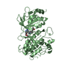
- PDB-4dcm: Crystal Structure of methyltransferase RlmG modifying G1835 of 23... -
+ Open data

- Basic information
| Entry | Database: PDB / ID: 4dcm | ||||||
|---|---|---|---|---|---|---|---|
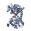
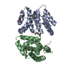
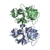
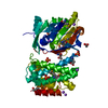
| Title | Crystal Structure of methyltransferase RlmG modifying G1835 of 23S rRNA in Escherichia coli | ||||||
 Components Components | Ribosomal RNA large subunit methyltransferase G | ||||||
 Keywords Keywords | TRANSFERASE / 23S rRNA (guanine1835-N2)-methyltransferase | ||||||
| Function / homology |  Function and homology information Function and homology information23S rRNA (guanine1835-N2)-methyltransferase / 23S rRNA (guanine(1835)-N(2))-methyltransferase activity / rRNA (guanine-N2-)-methyltransferase activity / rRNA base methylation / nucleic acid binding / cytoplasm Similarity search - Function | ||||||
| Biological species |  | ||||||
| Method |  X-RAY DIFFRACTION / SYNCHROTRON / SAD / Resolution: 2.297 Å X-RAY DIFFRACTION / SYNCHROTRON / SAD / Resolution: 2.297 Å | ||||||
 Authors Authors | Zhang, H. / Gao, Z.Q. / Dong, Y.H. | ||||||
 Citation Citation | Journal: Rna / Year: 2012 Title: Structural insights into the function of 23S rRNA methyltransferase RlmG (m2G1835) from Escherichia coli. Authors: Zhang, H. / Gao, Z.Q. / Wei, Y. / Wang, W.J. / Liu, G.F. / Shtykova, E.V. / Xu, J.H. / Dong, Y.H. | ||||||
| History |
|
- Structure visualization
| Structure viewer | Molecule: MolmilJmol/JSmol |
|---|
- Downloads & links
-Download
| PDBx/mmCIF format | 4dcm.cif.gz | 158.3 KB | Display | PDBx/mmCIF format |
|---|---|---|---|---|
| PDB format | pdb4dcm.ent.gz | 123.8 KB | Display | PDB format |
| PDBx/mmJSON format | 4dcm.json.gz | Tree view | PDBx/mmJSON format | |
| Others |  Other downloads Other downloads |
-Validation report
| Arichive directory | https://data.pdbj.org/pub/pdb/validation_reports/dc/4dcmftp://data.pdbj.org/pub/pdb/validation_reports/dc/4dcm | HTTPS FTP |
|---|
-Related structure data
| Similar structure data |
|---|


-Links
 PDBj
PDBj
- Assembly
| Deposited unit | 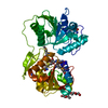
| ||||||||
|---|---|---|---|---|---|---|---|---|---|
| 1 |
| ||||||||
| Unit cell |
|
-Components
| #1: Protein | Mass: 42460.395 Da / Num. of mol.: 1 / Fragment: UNP RESIDUES 9-374 Source method: isolated from a genetically manipulated source Source: (gene. exp.) References: UniProt: P42596, 23S rRNA (guanine1835-N2)-methyltransferase | ||||
|---|---|---|---|---|---|
| #2: Chemical | ChemComp-SAM /   Mass: 398.437 Da / Num. of mol.: 1 / Source method: obtained synthetically / Formula: C15H22N6O5S Mass: 398.437 Da / Num. of mol.: 1 / Source method: obtained synthetically / Formula: C15H22N6O5S | ||||
| #3: Chemical |   Mass: 106.120 Da / Num. of mol.: 2 / Source method: obtained synthetically / Formula: C4H10O3 Mass: 106.120 Da / Num. of mol.: 2 / Source method: obtained synthetically / Formula: C4H10O3#4: Water | ChemComp-HOH / |  Mass: 18.015 Da / Num. of mol.: 128 / Source method: isolated from a natural source / Formula: H2O Mass: 18.015 Da / Num. of mol.: 128 / Source method: isolated from a natural source / Formula: H2OHas protein modification | Y | |
-Experimental details
-Experiment
| Experiment | Method: X-RAY DIFFRACTION / Number of used crystals: 1 |
|---|
- Sample preparation
| Crystal | Density Matthews: 2.34 Å3/Da / Density % sol: 47.39 % Description: THE ENTRY CONTAINS FRIEDEL PAIRS IN F_PLUS/MINUS COLUMNS |
|---|---|
| Crystal grow | Temperature: 300 K / Method: vapor diffusion, sitting drop / pH: 7.5 Details: 0.2M Tris pH7.5, 5% (w/v) PEG 8000, 1% (w/v) protamine sulfate, 0.02M HEPES sodium pH6.8, VAPOR DIFFUSION, SITTING DROP, temperature 300K |
-Data collection
| Diffraction | Mean temperature: 100 K |
|---|---|
| Diffraction source | Source: SYNCHROTRON / Site: BSRF  / Beamline: 3W1A / Wavelength: 0.9794 Å / Beamline: 3W1A / Wavelength: 0.9794 Å |
| Detector | Type: MAR CCD 165 mm / Detector: CCD / Date: Oct 31, 2011 |
| Radiation | Monochromator: double crystal monochromator / Protocol: SINGLE WAVELENGTH / Monochromatic (M) / Laue (L): M / Scattering type: x-ray |
| Radiation wavelength | Wavelength: 0.9794 Å / Relative weight: 1 |
| Reflection | Resolution: 2.297→50 Å / Num. all: 17821 / Num. obs: 16968 / % possible obs: 95.2 % / Observed criterion σ(F): 0 / Observed criterion σ(I): 0 / Redundancy: 7.2 % / Rmerge(I) obs: 0.06 / Net I/σ(I): 39.71 |
| Reflection shell | Resolution: 2.3→2.34 Å / Redundancy: 7 % / Rmerge(I) obs: 0.53 / Mean I/σ(I) obs: 5.62 / Num. unique all: 914 / % possible all: 100 |
- Processing
| Software |
| ||||||||||||||||||||||||||||||||||||||||||||||||||||||||||||||||||||||||||||||
|---|---|---|---|---|---|---|---|---|---|---|---|---|---|---|---|---|---|---|---|---|---|---|---|---|---|---|---|---|---|---|---|---|---|---|---|---|---|---|---|---|---|---|---|---|---|---|---|---|---|---|---|---|---|---|---|---|---|---|---|---|---|---|---|---|---|---|---|---|---|---|---|---|---|---|---|---|---|---|---|
| Refinement | Method to determine structure: SAD / Resolution: 2.297→29.984 Å / SU ML: 0.65 / σ(F): 1.34 / Phase error: 25.46 / Stereochemistry target values: ML Details: THE ENTRY CONTAINS FRIEDEL PAIRS IN F_PLUS/MINUS COLUMNS
| ||||||||||||||||||||||||||||||||||||||||||||||||||||||||||||||||||||||||||||||
| Solvent computation | Shrinkage radii: 0.83 Å / VDW probe radii: 1.1 Å / Solvent model: FLAT BULK SOLVENT MODEL / Bsol: 31.475 Å2 / ksol: 0.348 e/Å3 | ||||||||||||||||||||||||||||||||||||||||||||||||||||||||||||||||||||||||||||||
| Displacement parameters |
| ||||||||||||||||||||||||||||||||||||||||||||||||||||||||||||||||||||||||||||||
| Refinement step | Cycle: LAST / Resolution: 2.297→29.984 Å
| ||||||||||||||||||||||||||||||||||||||||||||||||||||||||||||||||||||||||||||||
| Refine LS restraints |
| ||||||||||||||||||||||||||||||||||||||||||||||||||||||||||||||||||||||||||||||
| LS refinement shell | Refine-ID: X-RAY DIFFRACTION / Total num. of bins used: 12
| ||||||||||||||||||||||||||||||||||||||||||||||||||||||||||||||||||||||||||||||
| Refinement TLS params. | Method: refined / Origin x: 36.9457 Å / Origin y: 1.1584 Å / Origin z: 54.2537 Å
| ||||||||||||||||||||||||||||||||||||||||||||||||||||||||||||||||||||||||||||||
| Refinement TLS group | Selection details: all |
