| 登録情報 | データベース: PDB / ID: 4dat
|
|---|
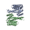
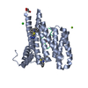
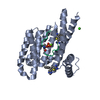
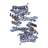
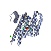
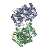
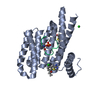
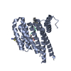
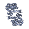
| タイトル | Structure of 14-3-3 sigma in complex with PADI6 14-3-3 binding motif II |
|---|
 要素 要素 | - 14-3-3 protein sigma
- Peptidylarginine Deiminase type VI
|
|---|
 キーワード キーワード | SIGNALING PROTEIN/PROTEIN BINDING / 14-3-3 fold / protein-protein interaction / SIGNALING PROTEIN-PROTEIN BINDING complex |
|---|
| 機能・相同性 |  機能・相同性情報 機能・相同性情報
regulation of translation by machinery localization / protein storage / structural constituent of cytoplasmic lattice / cytoplasmic lattice / cytoplasm organization / cortical granule / embryonic cleavage / intermediate filament cytoskeleton / epigenetic programming in the zygotic pronuclei / regulation of epidermal cell division ...regulation of translation by machinery localization / protein storage / structural constituent of cytoplasmic lattice / cytoplasmic lattice / cytoplasm organization / cortical granule / embryonic cleavage / intermediate filament cytoskeleton / epigenetic programming in the zygotic pronuclei / regulation of epidermal cell division / protein kinase C inhibitor activity / positive regulation of epidermal cell differentiation / keratinocyte development / keratinization / regulation of cell-cell adhesion / establishment of skin barrier / Regulation of localization of FOXO transcription factors / keratinocyte proliferation / Activation of BAD and translocation to mitochondria / phosphoserine residue binding / negative regulation of keratinocyte proliferation / cAMP/PKA signal transduction / negative regulation of protein localization to plasma membrane / SARS-CoV-2 targets host intracellular signalling and regulatory pathways / negative regulation of protein kinase activity / negative regulation of stem cell proliferation / SARS-CoV-1 targets host intracellular signalling and regulatory pathways / RHO GTPases activate PKNs / Chk1/Chk2(Cds1) mediated inactivation of Cyclin B:Cdk1 complex / Chromatin modifying enzymes / positive regulation of protein localization / cytoskeleton organization / positive regulation of cell adhesion / protein sequestering activity / negative regulation of innate immune response / TP53 Regulates Transcription of Genes Involved in G2 Cell Cycle Arrest / tubulin binding / protein export from nucleus / release of cytochrome c from mitochondria / positive regulation of protein export from nucleus / stem cell proliferation / TP53 Regulates Metabolic Genes / Translocation of SLC2A4 (GLUT4) to the plasma membrane / intrinsic apoptotic signaling pathway in response to DNA damage / intracellular protein localization / sperm midpiece / regulation of protein localization / positive regulation of cell growth / in utero embryonic development / regulation of cell cycle / cadherin binding / calcium ion binding / protein kinase binding / negative regulation of transcription by RNA polymerase II / signal transduction / extracellular space / extracellular exosome / identical protein binding / nucleus / cytoplasm / cytosol類似検索 - 分子機能 Protein-arginine deiminase / Protein-arginine deiminase, C-terminal / Protein-arginine deiminase (PAD), N-terminal / Protein-arginine deiminase (PAD), central domain / Protein-arginine deiminase, central domain superfamily / PAD, N-terminal domain superfamily / Protein-arginine deiminase (PAD) / Protein-arginine deiminase (PAD) N-terminal domain / Protein-arginine deiminase (PAD) middle domain / 14-3-3 domain ...Protein-arginine deiminase / Protein-arginine deiminase, C-terminal / Protein-arginine deiminase (PAD), N-terminal / Protein-arginine deiminase (PAD), central domain / Protein-arginine deiminase, central domain superfamily / PAD, N-terminal domain superfamily / Protein-arginine deiminase (PAD) / Protein-arginine deiminase (PAD) N-terminal domain / Protein-arginine deiminase (PAD) middle domain / 14-3-3 domain / Delta-Endotoxin; domain 1 / 14-3-3 protein sigma / 14-3-3 proteins signature 2. / 14-3-3 protein, conserved site / 14-3-3 proteins signature 1. / 14-3-3 protein / 14-3-3 homologues / 14-3-3 domain / 14-3-3 domain superfamily / 14-3-3 protein / Cupredoxin / Up-down Bundle / Mainly Alpha類似検索 - ドメイン・相同性 14-3-3 protein sigma / Inactive protein-arginine deiminase type-6 / Inactive protein-arginine deiminase type-6類似検索 - 構成要素 |
|---|
| 生物種 |  Homo sapiens (ヒト) Homo sapiens (ヒト) |
|---|
| 手法 |  X線回折 / シンクロトロン / 分子置換 / 解像度: 1.4 Å X線回折 / シンクロトロン / 分子置換 / 解像度: 1.4 Å |
|---|
 データ登録者 データ登録者 | Rose, R. / Rose, M. / Ottmann, C. |
|---|
 引用 引用 | ジャーナル: J.Struct.Biol. / 年: 2012
タイトル: Identification and structural characterization of two 14-3-3 binding sites in the human peptidylarginine deiminase type VI.
著者: Rose, R. / Rose, M. / Ottmann, C. |
|---|
| 履歴 | | 登録 | 2012年1月13日 | 登録サイト: RCSB / 処理サイト: RCSB |
|---|
| 改定 1.0 | 2012年6月13日 | Provider: repository / タイプ: Initial release |
|---|
| 改定 1.1 | 2012年10月24日 | Group: Database references |
|---|
| 改定 1.2 | 2024年10月30日 | Group: Data collection / Database references ...Data collection / Database references / Derived calculations / Structure summary
カテゴリ: chem_comp_atom / chem_comp_bond ...chem_comp_atom / chem_comp_bond / database_2 / pdbx_entry_details / pdbx_modification_feature / pdbx_struct_conn_angle / struct_conn / struct_ref_seq_dif / struct_site
Item: _database_2.pdbx_DOI / _database_2.pdbx_database_accession ..._database_2.pdbx_DOI / _database_2.pdbx_database_accession / _pdbx_struct_conn_angle.ptnr1_auth_seq_id / _pdbx_struct_conn_angle.ptnr1_label_alt_id / _pdbx_struct_conn_angle.ptnr1_label_atom_id / _pdbx_struct_conn_angle.ptnr1_label_seq_id / _pdbx_struct_conn_angle.ptnr3_auth_seq_id / _pdbx_struct_conn_angle.ptnr3_label_alt_id / _pdbx_struct_conn_angle.ptnr3_label_atom_id / _pdbx_struct_conn_angle.ptnr3_label_seq_id / _pdbx_struct_conn_angle.value / _struct_conn.pdbx_dist_value / _struct_conn.pdbx_leaving_atom_flag / _struct_conn.pdbx_ptnr1_label_alt_id / _struct_conn.ptnr1_auth_seq_id / _struct_conn.ptnr1_label_atom_id / _struct_conn.ptnr1_label_seq_id / _struct_conn.ptnr2_auth_seq_id / _struct_ref_seq_dif.details / _struct_site.pdbx_auth_asym_id / _struct_site.pdbx_auth_comp_id / _struct_site.pdbx_auth_seq_id |
|---|
|
|---|
 ムービー
ムービー コントローラー


 データを開く
 基本情報
基本情報 構造の表示
構造の表示 ダウンロードとリンク
 その他のダウンロード
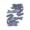


 PDBj
PDBj


 集合体
集合体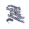


 分子量: 24.305 Da / 分子数: 1 / 由来タイプ: 合成 / 式: Mg
 分子量: 18.015 Da / 分子数: 393 / 由来タイプ: 天然 / 式: H2O
 試料調製
試料調製 / ビームライン: X10SA / 波長: 1.04 Å
 解析
解析