 Movie
Movie Controller

[English] 日本語
 Yorodumi
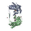
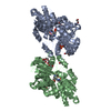
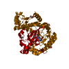
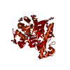
- PDB-4d9n: Crystal structure of Diaminopropionate ammonia lyase from Escheri... -
+ Open data

- Basic information
| Entry | Database: PDB / ID: 4d9n | ||||||
|---|---|---|---|---|---|---|---|
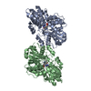
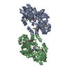
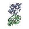
| Title | Crystal structure of Diaminopropionate ammonia lyase from Escherichia coli in complex with D-serine | ||||||
 Components Components | Diaminopropionate ammonia-lyase | ||||||
 Keywords Keywords | LYASE / Fold type II PLP-dependent enzyme / Tryptophan synthase beta subunit-like PLP-dependent enzymes superfamily | ||||||
| Function / homology |  Function and homology information Function and homology informationdiaminopropionate ammonia-lyase / diaminopropionate ammonia-lyase activity / pyridoxal phosphate binding / protein homodimerization activity Similarity search - Function | ||||||
| Biological species |  | ||||||
| Method |  X-RAY DIFFRACTION / MOLECULAR REPLACEMENT / Resolution: 2.5 Å X-RAY DIFFRACTION / MOLECULAR REPLACEMENT / Resolution: 2.5 Å | ||||||
 Authors Authors | Bisht, S. / Rajaram, V. / Bharath, S.R. / Murthy, M.R.N. | ||||||
 Citation Citation | Journal: J.Biol.Chem. / Year: 2012 Title: Crystal Structure of Escherichia coli Diaminopropionate Ammonia-lyase Reveals Mechanism of Enzyme Activation and Catalysis Authors: Bisht, S. / Rajaram, V. / Bharath, S.R. / Kalyani, J.N. / Khan, F. / Rao, A.N. / Savithri, H.S. / Murthy, M.R.N. | ||||||
| History |
|
- Structure visualization
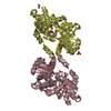
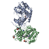
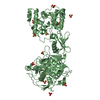
| Structure viewer | Molecule: MolmilJmol/JSmol |
|---|
- Downloads & links
-Download
| PDBx/mmCIF format | 4d9n.cif.gz | 304.2 KB | Display | PDBx/mmCIF format |
|---|---|---|---|---|
| PDB format | pdb4d9n.ent.gz | 249.6 KB | Display | PDB format |
| PDBx/mmJSON format | 4d9n.json.gz | Tree view | PDBx/mmJSON format | |
| Others |  Other downloads Other downloads |
-Validation report
| Summary document | 4d9n_validation.pdf.gz | 459.4 KB | Display | wwPDB validaton report |
|---|---|---|---|---|
| Full document | 4d9n_full_validation.pdf.gz | 463.4 KB | Display | |
| Data in XML | 4d9n_validation.xml.gz | 29.6 KB | Display | |
| Data in CIF | 4d9n_validation.cif.gz | 40 KB | Display | |
| Arichive directory | https://data.pdbj.org/pub/pdb/validation_reports/d9/4d9nftp://data.pdbj.org/pub/pdb/validation_reports/d9/4d9n | HTTPS FTP |
-Related structure data
| Related structure data |  4d9gC  4d9iSC 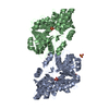 4d9kC  4d9mC C: citing same article ( S: Starting model for refinement |
|---|---|
| Similar structure data |
-Links
 PDBj
PDBj
- Assembly
| Deposited unit | 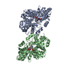
| ||||||||
|---|---|---|---|---|---|---|---|---|---|
| 1 |
| ||||||||
| Unit cell |
|
-Components
| #1: Protein | Mass: 43600.535 Da / Num. of mol.: 2 Source method: isolated from a genetically manipulated source Source: (gene. exp.) References: UniProt: P66899, diaminopropionate ammonia-lyase #2: Chemical | 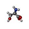  Type: D-peptide linking / Mass: 105.093 Da / Num. of mol.: 2 / Source method: obtained synthetically / Formula: C3H7NO3 Type: D-peptide linking / Mass: 105.093 Da / Num. of mol.: 2 / Source method: obtained synthetically / Formula: C3H7NO3#3: Water | ChemComp-HOH / |  Mass: 18.015 Da / Num. of mol.: 101 / Source method: isolated from a natural source / Formula: H2O Mass: 18.015 Da / Num. of mol.: 101 / Source method: isolated from a natural source / Formula: H2O |
|---|
-Experimental details
-Experiment
| Experiment | Method: X-RAY DIFFRACTION / Number of used crystals: 1 |
|---|
- Sample preparation
| Crystal | Density Matthews: 2.18 Å3/Da / Density % sol: 43.62 % |
|---|---|
| Crystal grow | Temperature: 298 K / Method: vapor diffusion, hanging drop / pH: 8.3 Details: 20% PEG 3350, 70mM magnesium chloride, 50mM lithium sulfate, 6mM sodium citrate, 40mM tris HCl pH 8.3. Crystals were soaked with 10mM D-ser in crystallization condition for 10 hrs before ...Details: 20% PEG 3350, 70mM magnesium chloride, 50mM lithium sulfate, 6mM sodium citrate, 40mM tris HCl pH 8.3. Crystals were soaked with 10mM D-ser in crystallization condition for 10 hrs before data collection., VAPOR DIFFUSION, HANGING DROP, temperature 298K |
-Data collection
| Diffraction | Mean temperature: 100 K |
|---|---|
| Diffraction source | Source: ROTATING ANODE / Type: BRUKER AXS MICROSTAR / Wavelength: 1.54179 Å |
| Detector | Type: MAR scanner 345 mm plate / Detector: IMAGE PLATE / Date: Nov 29, 2010 / Details: mirror |
| Radiation | Monochromator: osmic mirror / Protocol: SINGLE WAVELENGTH / Monochromatic (M) / Laue (L): M / Scattering type: x-ray |
| Radiation wavelength | Wavelength: 1.54179 Å / Relative weight: 1 |
| Reflection | Resolution: 2.5→50 Å / Num. obs: 27635 / % possible obs: 99.8 % / Redundancy: 9.5 % / Biso Wilson estimate: 49.3 Å2 / Rmerge(I) obs: 0.114 / Net I/σ(I): 31.2 |
| Reflection shell | Resolution: 2.5→2.54 Å / Redundancy: 5 % / Rmerge(I) obs: 0.389 / Mean I/σ(I) obs: 3.6 / Num. unique all: 1311 / % possible all: 97.3 |
- Processing
| Software |
| |||||||||||||||||||||||||||||||||||||||||||||||||||||||||||||||||||||||||||||||||||||||||||||||||||||||||||||||||||||||||||||||||||||||||||||||||||||||||||||||||||||||||||||||||||||||||||||||||||||||||||||||||||||||||||||||||
|---|---|---|---|---|---|---|---|---|---|---|---|---|---|---|---|---|---|---|---|---|---|---|---|---|---|---|---|---|---|---|---|---|---|---|---|---|---|---|---|---|---|---|---|---|---|---|---|---|---|---|---|---|---|---|---|---|---|---|---|---|---|---|---|---|---|---|---|---|---|---|---|---|---|---|---|---|---|---|---|---|---|---|---|---|---|---|---|---|---|---|---|---|---|---|---|---|---|---|---|---|---|---|---|---|---|---|---|---|---|---|---|---|---|---|---|---|---|---|---|---|---|---|---|---|---|---|---|---|---|---|---|---|---|---|---|---|---|---|---|---|---|---|---|---|---|---|---|---|---|---|---|---|---|---|---|---|---|---|---|---|---|---|---|---|---|---|---|---|---|---|---|---|---|---|---|---|---|---|---|---|---|---|---|---|---|---|---|---|---|---|---|---|---|---|---|---|---|---|---|---|---|---|---|---|---|---|---|---|---|---|---|---|---|---|---|---|---|---|---|---|---|---|---|---|---|---|
| Refinement | Method to determine structure: MOLECULAR REPLACEMENT Starting model: PDB ENTRY 4D9I Resolution: 2.5→45.57 Å / Cor.coef. Fo:Fc: 0.93 / Cor.coef. Fo:Fc free: 0.887 / SU B: 22.865 / SU ML: 0.255 / Cross valid method: THROUGHOUT / ESU R: 0.977 / ESU R Free: 0.328 / Stereochemistry target values: MAXIMUM LIKELIHOOD / Details: HYDROGENS HAVE BEEN USED IF PRESENT IN THE INPUT
| |||||||||||||||||||||||||||||||||||||||||||||||||||||||||||||||||||||||||||||||||||||||||||||||||||||||||||||||||||||||||||||||||||||||||||||||||||||||||||||||||||||||||||||||||||||||||||||||||||||||||||||||||||||||||||||||||
| Solvent computation | Ion probe radii: 0.8 Å / Shrinkage radii: 0.8 Å / VDW probe radii: 1.2 Å / Solvent model: MASK | |||||||||||||||||||||||||||||||||||||||||||||||||||||||||||||||||||||||||||||||||||||||||||||||||||||||||||||||||||||||||||||||||||||||||||||||||||||||||||||||||||||||||||||||||||||||||||||||||||||||||||||||||||||||||||||||||
| Displacement parameters | Biso mean: 43.121 Å2
| |||||||||||||||||||||||||||||||||||||||||||||||||||||||||||||||||||||||||||||||||||||||||||||||||||||||||||||||||||||||||||||||||||||||||||||||||||||||||||||||||||||||||||||||||||||||||||||||||||||||||||||||||||||||||||||||||
| Refinement step | Cycle: LAST / Resolution: 2.5→45.57 Å
| |||||||||||||||||||||||||||||||||||||||||||||||||||||||||||||||||||||||||||||||||||||||||||||||||||||||||||||||||||||||||||||||||||||||||||||||||||||||||||||||||||||||||||||||||||||||||||||||||||||||||||||||||||||||||||||||||
| Refine LS restraints |
| |||||||||||||||||||||||||||||||||||||||||||||||||||||||||||||||||||||||||||||||||||||||||||||||||||||||||||||||||||||||||||||||||||||||||||||||||||||||||||||||||||||||||||||||||||||||||||||||||||||||||||||||||||||||||||||||||
| LS refinement shell | Resolution: 2.5→2.564 Å / Total num. of bins used: 20
| |||||||||||||||||||||||||||||||||||||||||||||||||||||||||||||||||||||||||||||||||||||||||||||||||||||||||||||||||||||||||||||||||||||||||||||||||||||||||||||||||||||||||||||||||||||||||||||||||||||||||||||||||||||||||||||||||
| Refinement TLS params. | Method: refined / Refine-ID: X-RAY DIFFRACTION
| |||||||||||||||||||||||||||||||||||||||||||||||||||||||||||||||||||||||||||||||||||||||||||||||||||||||||||||||||||||||||||||||||||||||||||||||||||||||||||||||||||||||||||||||||||||||||||||||||||||||||||||||||||||||||||||||||
| Refinement TLS group |
|
