 Movie
Movie Controller

[English] 日本語
 Yorodumi
- PDB-4d7c: Monoclinic crystal form of the extracellular olfactomedin domain ... -
+ Open data

- Basic information
| Entry | Database: PDB / ID: 4d7c | ||||||
|---|---|---|---|---|---|---|---|
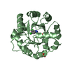
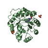
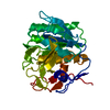
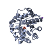
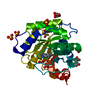
| Title | Monoclinic crystal form of the extracellular olfactomedin domain from gliomedin | ||||||
 Components Components | GLIOMEDIN | ||||||
 Keywords Keywords | SIGNALING PROTEIN / MYELIN / BETA-PROPELLER | ||||||
| Function / homology |  Function and homology information Function and homology informationclustering of voltage-gated sodium channels / protein binding involved in heterotypic cell-cell adhesion / microvillus organization / axon / cell surface / signal transduction / : / plasma membrane Similarity search - Function | ||||||
| Biological species |  | ||||||
| Method |  X-RAY DIFFRACTION / SYNCHROTRON / MOLECULAR REPLACEMENT / Resolution: 1.45 Å X-RAY DIFFRACTION / SYNCHROTRON / MOLECULAR REPLACEMENT / Resolution: 1.45 Å | ||||||
 Authors Authors | Han, H. / Kursula, P. | ||||||
 Citation Citation | Journal: J.Biol.Chem. / Year: 2015 Title: The Olfactomedin Domain from Gliomedin is a Beta-Propeller with Unique Structural Properties. Authors: Han, H. / Kursula, P. | ||||||
| History |
|
- Structure visualization
| Structure viewer | Molecule: MolmilJmol/JSmol |
|---|
- Downloads & links
-Download
| PDBx/mmCIF format | 4d7c.cif.gz | 321.3 KB | Display | PDBx/mmCIF format |
|---|---|---|---|---|
| PDB format | pdb4d7c.ent.gz | 266.9 KB | Display | PDB format |
| PDBx/mmJSON format | 4d7c.json.gz | Tree view | PDBx/mmJSON format | |
| Others |  Other downloads Other downloads |
-Validation report
| Arichive directory | https://data.pdbj.org/pub/pdb/validation_reports/d7/4d7cftp://data.pdbj.org/pub/pdb/validation_reports/d7/4d7c | HTTPS FTP |
|---|
-Related structure data
| Related structure data |  4d77SC S: Starting model for refinement C: citing same article ( |
|---|---|
| Similar structure data |


-Links
 PDBj
PDBj- Assembly
| Deposited unit | 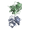
| ||||||||
|---|---|---|---|---|---|---|---|---|---|
| 1 | 
| ||||||||
| 2 | 
| ||||||||
| Unit cell |
|
-Components
| #1: Protein | Mass: 32435.500 Da / Num. of mol.: 2 / Fragment: OLFACTOMEDIN DOMAIN, UNP RESIDUES 260-543 Source method: isolated from a genetically manipulated source Source: (gene. exp.)   SPODOPTERA FRUGIPERDA (fall armyworm) / References: UniProt: Q80WL1 SPODOPTERA FRUGIPERDA (fall armyworm) / References: UniProt: Q80WL1#2: Chemical |   Mass: 22.990 Da / Num. of mol.: 2 / Source method: obtained synthetically / Formula: Na Mass: 22.990 Da / Num. of mol.: 2 / Source method: obtained synthetically / Formula: Na#3: Water | ChemComp-HOH / |  Mass: 18.015 Da / Num. of mol.: 561 / Source method: isolated from a natural source / Formula: H2O Mass: 18.015 Da / Num. of mol.: 561 / Source method: isolated from a natural source / Formula: H2O |
|---|
-Experimental details
-Experiment
| Experiment | Method: X-RAY DIFFRACTION / Number of used crystals: 1 |
|---|
- Sample preparation
| Crystal | Density % sol: 40 % / Description: NONE |
|---|---|
| Crystal grow | Details: 100 MM SODIUM CITRATE PH 5.5, 20% PEG 3000 |
-Data collection
| Diffraction | Mean temperature: 100 K |
|---|---|
| Diffraction source | Source: SYNCHROTRON / Site: ESRF  / Beamline: ID23-2 / Wavelength: 0.873 / Beamline: ID23-2 / Wavelength: 0.873 |
| Detector | Type: ADSC CCD / Detector: CCD |
| Radiation | Protocol: SINGLE WAVELENGTH / Monochromatic (M) / Laue (L): M / Scattering type: x-ray |
| Radiation wavelength | Wavelength: 0.873 Å / Relative weight: 1 |
| Reflection | Resolution: 1.45→50 Å / Num. obs: 78451 / % possible obs: 98.7 % / Observed criterion σ(I): -3 / Redundancy: 3.7 % / Biso Wilson estimate: 12.99 Å2 / Rmerge(I) obs: 0.1 / Net I/σ(I): 8.4 |
| Reflection shell | Resolution: 1.45→1.49 Å / Redundancy: 3.6 % / Rmerge(I) obs: 1.02 / Mean I/σ(I) obs: 1.3 / % possible all: 97.4 |
- Processing
| Software |
| ||||||||||||||||||||||||||||||||||||||||||||||||||||||||||||||||||||||||||||||||||||
|---|---|---|---|---|---|---|---|---|---|---|---|---|---|---|---|---|---|---|---|---|---|---|---|---|---|---|---|---|---|---|---|---|---|---|---|---|---|---|---|---|---|---|---|---|---|---|---|---|---|---|---|---|---|---|---|---|---|---|---|---|---|---|---|---|---|---|---|---|---|---|---|---|---|---|---|---|---|---|---|---|---|---|---|---|---|
| Refinement | Method to determine structure: MOLECULAR REPLACEMENT Starting model: PDB ENTRY 4D77 Resolution: 1.45→43.084 Å / SU ML: 0.18 / σ(F): 1.36 / Phase error: 24.2 / Stereochemistry target values: ML
| ||||||||||||||||||||||||||||||||||||||||||||||||||||||||||||||||||||||||||||||||||||
| Solvent computation | Shrinkage radii: 0.9 Å / VDW probe radii: 1.11 Å / Solvent model: FLAT BULK SOLVENT MODEL | ||||||||||||||||||||||||||||||||||||||||||||||||||||||||||||||||||||||||||||||||||||
| Refinement step | Cycle: LAST / Resolution: 1.45→43.084 Å
| ||||||||||||||||||||||||||||||||||||||||||||||||||||||||||||||||||||||||||||||||||||
| Refine LS restraints |
| ||||||||||||||||||||||||||||||||||||||||||||||||||||||||||||||||||||||||||||||||||||
| LS refinement shell |
|
