 Movie
Movie Controller

[English] 日本語
 Yorodumi
- PDB-4d0u: Crystal structure of the fiber head domain of the Atadenovirus sn... -
+ Open data

- Basic information
| Entry | Database: PDB / ID: 4d0u | |||||||||
|---|---|---|---|---|---|---|---|---|---|---|
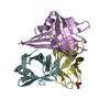
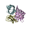
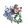
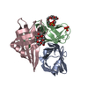
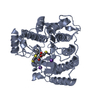
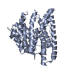
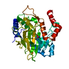
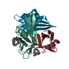
| Title | Crystal structure of the fiber head domain of the Atadenovirus snake adenovirus 1, selenomethionine-derivative | |||||||||
 Components Components | FIBER PROTEIN | |||||||||
 Keywords Keywords | VIRAL PROTEIN | |||||||||
| Function / homology |  Function and homology information Function and homology informationviral capsid / cell adhesion / symbiont entry into host cell / virion attachment to host cell / host cell nucleus Similarity search - Function | |||||||||
| Biological species |  SNAKE ADENOVIRUS 1 SNAKE ADENOVIRUS 1 | |||||||||
| Method |  X-RAY DIFFRACTION / SYNCHROTRON / MAD / Resolution: 1.6 Å X-RAY DIFFRACTION / SYNCHROTRON / MAD / Resolution: 1.6 Å | |||||||||
 Authors Authors | Singh, A.K. / van Raaij, M.J. | |||||||||
 Citation Citation | Journal: PLoS ONE / Year: 2014 Title: Crystal structure of the fibre head domain of the Atadenovirus Snake Adenovirus 1. Authors: Singh, A.K. / Menendez-Conejero, R. / San Martin, C. / van Raaij, M.J. #1: Journal: Acta Crystallogr. Sect. F Struct. Biol. Cryst. Commun. Year: 2013 Title: Crystallization of the C-terminal domain of the fibre protein from snake adenovirus 1, an atadenovirus. Authors: Singh, A.K. / Menendez-Conejero, R. / San Martin, C. / van Raaij, M.J. #2: Journal: Archives of Virology / Year: 1993 Title: Physicochemical Properties and Cytopathogenicity of an Adenovirus-Like Agent Isolated from Corn Snake (Elaphe Guttata). Authors: Juhasz, A. / Ahne, W. #3: Journal: Virus Res. / Year: 2008 Title: Completion of the Genome Analysis of Snake Adenovirus Type 1, a Representative of the Reptilian Lineage within the Novel Genus Atadenovirus. Authors: Farkas, S.L. / Harrach, B. / Benko, M. | |||||||||
| History |
|
- Structure visualization
| Structure viewer | Molecule: MolmilJmol/JSmol |
|---|
- Downloads & links
-Download
| PDBx/mmCIF format | 4d0u.cif.gz | 112.9 KB | Display | PDBx/mmCIF format |
|---|---|---|---|---|
| PDB format | pdb4d0u.ent.gz | 88.2 KB | Display | PDB format |
| PDBx/mmJSON format | 4d0u.json.gz | Tree view | PDBx/mmJSON format | |
| Others |  Other downloads Other downloads |
-Validation report
| Arichive directory | https://data.pdbj.org/pub/pdb/validation_reports/d0/4d0uftp://data.pdbj.org/pub/pdb/validation_reports/d0/4d0u | HTTPS FTP |
|---|
-Related structure data


-Links
 PDBj
PDBj
- Assembly
| Deposited unit | 
| ||||||||||||||||||||||||||||||
|---|---|---|---|---|---|---|---|---|---|---|---|---|---|---|---|---|---|---|---|---|---|---|---|---|---|---|---|---|---|---|---|
| 1 | 
| ||||||||||||||||||||||||||||||
| 2 | 
| ||||||||||||||||||||||||||||||
| Unit cell |
| ||||||||||||||||||||||||||||||
| Components on special symmetry positions |
| ||||||||||||||||||||||||||||||
| Noncrystallographic symmetry (NCS) | NCS oper:
|
-Components
| #1: Protein | Mass: 15640.671 Da / Num. of mol.: 4 / Fragment: FIBER HEAD DOMAIN, RESIDUES 234-339 / Mutation: YES Source method: isolated from a genetically manipulated source Source: (gene. exp.) SNAKE ADENOVIRUS 1Description: SNAKE ADENOVIRUS 1 (SNADV1) WAS FIRST ISOLATED FROM THE CORN SNAKE ELAPHE GUTTATA, JUHASZ & AHNE, 1993, I.E. ADDITIONAL REFERENCE 2. Plasmid: PET28C / Production host:  #2: Chemical |   Mass: 370.436 Da / Num. of mol.: 2 / Source method: obtained synthetically / Formula: C16H34O9 Mass: 370.436 Da / Num. of mol.: 2 / Source method: obtained synthetically / Formula: C16H34O9#3: Chemical | ChemComp-SO4 /   Mass: 96.063 Da / Num. of mol.: 4 / Source method: obtained synthetically / Formula: SO4 Mass: 96.063 Da / Num. of mol.: 4 / Source method: obtained synthetically / Formula: SO4#4: Water | ChemComp-HOH / |  Mass: 18.015 Da / Num. of mol.: 534 / Source method: isolated from a natural source / Formula: H2O Mass: 18.015 Da / Num. of mol.: 534 / Source method: isolated from a natural source / Formula: H2OHas protein modification | Y | Sequence details | LEU322 AND LEU324 WERE MUTATED TO MET. THE C-TERMINAL PART OF THE SEQUENCE IS DIFFERENT, WE BELIEVE ...LEU322 AND LEU324 WERE MUTATED TO MET. THE C-TERMINAL PART OF THE SEQUENCE IS DIFFERENT, WE BELIEVE THE DATABASE SEQUENCE IS WRONG. | |
|---|
-Experimental details
-Experiment
| Experiment | Method: X-RAY DIFFRACTION / Number of used crystals: 1 |
|---|
- Sample preparation
| Crystal | Density Matthews: 2.24 Å3/Da / Density % sol: 45.24 % / Description: NONE |
|---|---|
| Crystal grow | pH: 7.5 Details: 10 MM TRIS-HCL PH 8.5, 10 MM BETA-MERCAPTOETHANOL, 1.7 M AMMONIUM SULFATE, 0.085 M HEPES SODIUM SALT PH 7.5, 1.7%(V/V) POLYETHYLENE GLYCOL (PEG) 400, 15%(V/V) GLYCEROL |
-Data collection
| Diffraction | Mean temperature: 100 K | ||||||||||||
|---|---|---|---|---|---|---|---|---|---|---|---|---|---|
| Diffraction source | Source: SYNCHROTRON / Site: ESRF  / Beamline: ID14-4 / Wavelength: 0.9768, 0.9791, 0.9793 / Beamline: ID14-4 / Wavelength: 0.9768, 0.9791, 0.9793 | ||||||||||||
| Detector | Type: ADSC QUANTUM 315r / Detector: CCD / Date: Oct 26, 2012 / Details: TOROIDAL FOCUSING MIRROR | ||||||||||||
| Radiation | Monochromator: CHANNEL CUT ESRF MONOCHROMATOR SI(111) / Protocol: MAD / Monochromatic (M) / Laue (L): M / Scattering type: x-ray | ||||||||||||
| Radiation wavelength |
| ||||||||||||
| Reflection | Resolution: 1.6→30 Å / Num. obs: 72960 / % possible obs: 100 % / Redundancy: 12.2 % / Biso Wilson estimate: 13.9 Å2 / Rmerge(I) obs: 0.09 / Net I/σ(I): 18.2 | ||||||||||||
| Reflection shell | Resolution: 1.6→1.69 Å / Redundancy: 12.2 % / Rmerge(I) obs: 0.4 / Mean I/σ(I) obs: 6.2 / % possible all: 100 |
- Processing
| Software |
| ||||||||||||||||||||||||||||||||||||||||||||||||||||||||||||||||||||||||||||||||||||||||||||||||||||||||||||||||||||||||||||||||||||||||||||||||||||||||||||||||||||||||||||||||||||||
|---|---|---|---|---|---|---|---|---|---|---|---|---|---|---|---|---|---|---|---|---|---|---|---|---|---|---|---|---|---|---|---|---|---|---|---|---|---|---|---|---|---|---|---|---|---|---|---|---|---|---|---|---|---|---|---|---|---|---|---|---|---|---|---|---|---|---|---|---|---|---|---|---|---|---|---|---|---|---|---|---|---|---|---|---|---|---|---|---|---|---|---|---|---|---|---|---|---|---|---|---|---|---|---|---|---|---|---|---|---|---|---|---|---|---|---|---|---|---|---|---|---|---|---|---|---|---|---|---|---|---|---|---|---|---|---|---|---|---|---|---|---|---|---|---|---|---|---|---|---|---|---|---|---|---|---|---|---|---|---|---|---|---|---|---|---|---|---|---|---|---|---|---|---|---|---|---|---|---|---|---|---|---|---|
| Refinement | Method to determine structure: MAD Starting model: NONE Resolution: 1.6→29.33 Å / Cor.coef. Fo:Fc: 0.968 / Cor.coef. Fo:Fc free: 0.95 / SU B: 1.324 / SU ML: 0.047 / Cross valid method: THROUGHOUT / ESU R: 0.068 / ESU R Free: 0.072 / Stereochemistry target values: MAXIMUM LIKELIHOOD Details: HYDROGENS HAVE BEEN ADDED IN THE RIDING POSITIONS. U VALUES REFINED INDIVIDUALLY
| ||||||||||||||||||||||||||||||||||||||||||||||||||||||||||||||||||||||||||||||||||||||||||||||||||||||||||||||||||||||||||||||||||||||||||||||||||||||||||||||||||||||||||||||||||||||
| Solvent computation | Ion probe radii: 0.8 Å / Shrinkage radii: 0.8 Å / VDW probe radii: 1.2 Å / Solvent model: MASK | ||||||||||||||||||||||||||||||||||||||||||||||||||||||||||||||||||||||||||||||||||||||||||||||||||||||||||||||||||||||||||||||||||||||||||||||||||||||||||||||||||||||||||||||||||||||
| Displacement parameters | Biso mean: 18.448 Å2 | ||||||||||||||||||||||||||||||||||||||||||||||||||||||||||||||||||||||||||||||||||||||||||||||||||||||||||||||||||||||||||||||||||||||||||||||||||||||||||||||||||||||||||||||||||||||
| Refinement step | Cycle: LAST / Resolution: 1.6→29.33 Å
| ||||||||||||||||||||||||||||||||||||||||||||||||||||||||||||||||||||||||||||||||||||||||||||||||||||||||||||||||||||||||||||||||||||||||||||||||||||||||||||||||||||||||||||||||||||||
| Refine LS restraints |
|