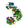
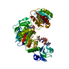
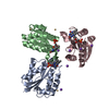
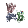
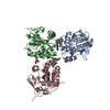
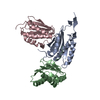
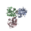
Entry Database : PDB / ID : 4cvkTitle PaMurF in complex with UDP-MurNAc-tripeptide (mDAP) ALA-FGA-API UDP-N-acetylmuramoyl-tripeptide--D-alanyl-D-alanine ligase Keywords / / Function / homology Function Domain/homology Component
/ / / / / / / / / / / / / / / / / / / / / / / / / / / / / / / / / / / / / Biological species Pseudomonas aeruginosa PAO1 (bacteria)synthetic construct (others) Method / / / Resolution : 1.92 Å Authors Ruane, K.M. / Majce, V. Journal : To be Published Title : Pamurf in Complex with Udp-Murnac-Tripeptide (Mdap)Authors : Majce, V. / Ruane, K. History Deposition Mar 28, 2014 Deposition site / Processing site Revision 1.0 Apr 22, 2015 Provider / Type Revision 1.1 Apr 29, 2015 Group Revision 1.2 Sep 23, 2015 Group Revision 1.3 Jul 29, 2020 Group Data collection / Derived calculations ... Data collection / Derived calculations / Other / Structure summary Category chem_comp / entity ... chem_comp / entity / pdbx_chem_comp_identifier / pdbx_database_status / pdbx_entity_nonpoly / struct_conn / struct_site / struct_site_gen Item _chem_comp.name / _chem_comp.type ... _chem_comp.name / _chem_comp.type / _entity.pdbx_description / _pdbx_database_status.status_code_sf / _pdbx_entity_nonpoly.name / _struct_conn.pdbx_leaving_atom_flag Description / Provider / Type Revision 2.0 Dec 7, 2022 Group Atomic model / Data collection ... Atomic model / Data collection / Database references / Derived calculations / Polymer sequence / Source and taxonomy / Structure summary Category atom_site / chem_comp ... atom_site / chem_comp / database_2 / entity / entity_poly / entity_poly_seq / entity_src_gen / pdbx_entity_nonpoly / pdbx_entity_src_syn / pdbx_nonpoly_scheme / pdbx_poly_seq_scheme / pdbx_struct_assembly / pdbx_struct_assembly_gen / pdbx_struct_assembly_prop / struct_asym / struct_conn / struct_ref / struct_ref_seq Item _atom_site.B_iso_or_equiv / _atom_site.Cartn_x ... _atom_site.B_iso_or_equiv / _atom_site.Cartn_x / _atom_site.Cartn_y / _atom_site.Cartn_z / _atom_site.auth_asym_id / _atom_site.auth_atom_id / _atom_site.auth_comp_id / _atom_site.auth_seq_id / _atom_site.group_PDB / _atom_site.label_asym_id / _atom_site.label_atom_id / _atom_site.label_comp_id / _atom_site.label_entity_id / _atom_site.label_seq_id / _atom_site.type_symbol / _chem_comp.pdbx_synonyms / _database_2.pdbx_DOI / _database_2.pdbx_database_accession / _entity_src_gen.pdbx_beg_seq_num / _entity_src_gen.pdbx_end_seq_num / _entity_src_gen.pdbx_gene_src_gene / _entity_src_gen.pdbx_gene_src_scientific_name / _entity_src_gen.pdbx_seq_type / _pdbx_struct_assembly.details / _pdbx_struct_assembly.oligomeric_count / _pdbx_struct_assembly.oligomeric_details / _pdbx_struct_assembly_gen.asym_id_list / _struct_conn.ptnr1_auth_asym_id / _struct_conn.ptnr1_label_asym_id / _struct_conn.ptnr1_label_seq_id / _struct_conn.ptnr2_auth_asym_id / _struct_conn.ptnr2_label_asym_id / _struct_conn.ptnr2_label_seq_id Revision 3.0 Nov 15, 2023 Group / Data collection / Derived calculationsCategory atom_site / chem_comp_atom ... atom_site / chem_comp_atom / chem_comp_bond / struct_conn Item _atom_site.auth_atom_id / _atom_site.label_atom_id ... _atom_site.auth_atom_id / _atom_site.label_atom_id / _struct_conn.pdbx_leaving_atom_flag / _struct_conn.ptnr2_label_atom_id Revision 3.1 Jan 31, 2024 Group / Category
Show all Show less
 Movie
Movie Controller


 Open data
 Basic information
 Components
 Keywords
 Function and homology information
 Pseudomonas aeruginosa PAO1 (bacteria)
 X-RAY DIFFRACTION /
  Authors
 Citation
 Structure visualization
 Downloads & links
 Other downloads
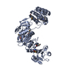


 PDBj
PDBj


 Assembly

 Type: RNA linking / Mass: 404.161 Da / Num. of mol.: 1 / Source method: obtained synthetically / Formula: C9H14N2O12P2 / Comment: UDP*YM
 Type: D-saccharide, alpha linking / Mass: 293.270 Da / Num. of mol.: 1 / Source method: obtained synthetically / Formula: C11H19NO8
 Mass: 18.015 Da / Num. of mol.: 84 / Source method: isolated from a natural source / Formula: H2O
 Sample preparation
 / Beamline: I04-1 / Wavelength: 0.92
  Processing