 Movie
Movie Controller

[English] 日本語
 Yorodumi
- PDB-4com: Crystal structure of the anaerobic ribonucleotide reductase from ... -
+ Open data

- Basic information
| Entry | Database: PDB / ID: 4com | |||||||||
|---|---|---|---|---|---|---|---|---|---|---|
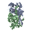
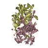
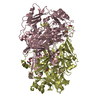
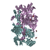
| Title | Crystal structure of the anaerobic ribonucleotide reductase from Thermotoga maritima with MES in the active site | |||||||||
 Components Components | ANAEROBIC RIBONUCLEOSIDE-TRIPHOSPHATE REDUCTASE | |||||||||
 Keywords Keywords | OXIDOREDUCTASE / RADICAL CHEMISTRY / ALLOSTERIC REGULATION / ANAEROBIC ENZYME | |||||||||
| Function / homology | anaerobic ribonucleoside-triphosphate reductase complex / Ribonucleoside-triphosphate reductase, anaerobic / Anaerobic ribonucleoside-triphosphate reductase / ribonucleoside-triphosphate reductase (thioredoxin) activity / 2'-deoxyribonucleotide biosynthetic process / DNA replication / nucleotide binding / metal ion binding / Uncharacterized protein Function and homology information Function and homology information | |||||||||
| Biological species |   THERMOTOGA MARITIMA (bacteria) THERMOTOGA MARITIMA (bacteria) | |||||||||
| Method |  X-RAY DIFFRACTION / SYNCHROTRON / MOLECULAR REPLACEMENT / Resolution: 1.92 Å X-RAY DIFFRACTION / SYNCHROTRON / MOLECULAR REPLACEMENT / Resolution: 1.92 Å | |||||||||
 Authors Authors | Aurelius, O. / Johansson, R. / Bagenholm, V. / Beck, T. / Balhuizen, A. / Lundin, D. / Sjoberg, B.M. / Mulliez, E. / Logan, D.T. | |||||||||
 Citation Citation | Journal: Plos One / Year: 2015 Title: The Crystal Structure of Thermotoga Maritima Class III Ribonucleotide Reductase Lacks a Radical Cysteine Pre-Positioned in the Active Site. Authors: Aurelius, O. / Johansson, R. / Bagenholm, V. / Beck, T. / Balhuizen, A. / Lundin, D. / Sjoberg, B.M. / Mulliez, E. / Logan, D.T. | |||||||||
| History |
|
- Structure visualization
| Structure viewer | Molecule: MolmilJmol/JSmol |
|---|
- Downloads & links
-Download
| PDBx/mmCIF format | 4com.cif.gz | 719.5 KB | Display | PDBx/mmCIF format |
|---|---|---|---|---|
| PDB format | pdb4com.ent.gz | 607.2 KB | Display | PDB format |
| PDBx/mmJSON format | 4com.json.gz | Tree view | PDBx/mmJSON format | |
| Others |  Other downloads Other downloads |
-Validation report
| Arichive directory | https://data.pdbj.org/pub/pdb/validation_reports/co/4comftp://data.pdbj.org/pub/pdb/validation_reports/co/4com | HTTPS FTP |
|---|
-Related structure data
| Related structure data |  4coiSC  4cojC  4colC  4conC S: Starting model for refinement C: citing same article ( |
|---|---|
| Similar structure data |


-Links
 PDBj
PDBj- Assembly
| Deposited unit | 
| ||||||||
|---|---|---|---|---|---|---|---|---|---|
| 1 |
| ||||||||
| Unit cell |
| ||||||||
| Noncrystallographic symmetry (NCS) | NCS oper: (Code: given Matrix: (0.6612, 0.02988, -0.7496), Vector: |
-Components
| #1: Protein | Mass: 75674.383 Da / Num. of mol.: 2 Source method: isolated from a genetically manipulated source Source: (gene. exp.) THERMOTOGA MARITIMA (bacteria) / Strain: MSB8 / Production host: References: UniProt: Q9WYL6, ribonucleoside-triphosphate reductase (thioredoxin) #2: Chemical |   Mass: 65.409 Da / Num. of mol.: 2 / Source method: obtained synthetically / Formula: Zn Mass: 65.409 Da / Num. of mol.: 2 / Source method: obtained synthetically / Formula: Zn#3: Chemical |   Mass: 195.237 Da / Num. of mol.: 2 / Source method: obtained synthetically / Formula: C6H13NO4S / Comment: pH buffer*YM Mass: 195.237 Da / Num. of mol.: 2 / Source method: obtained synthetically / Formula: C6H13NO4S / Comment: pH buffer*YM#4: Chemical | ChemComp-1PE / |   Mass: 238.278 Da / Num. of mol.: 1 / Source method: obtained synthetically / Formula: C10H22O6 / Comment: precipitant*YM Mass: 238.278 Da / Num. of mol.: 1 / Source method: obtained synthetically / Formula: C10H22O6 / Comment: precipitant*YM#5: Water | ChemComp-HOH / |  Mass: 18.015 Da / Num. of mol.: 712 / Source method: isolated from a natural source / Formula: H2O Mass: 18.015 Da / Num. of mol.: 712 / Source method: isolated from a natural source / Formula: H2O |
|---|
-Experimental details
-Experiment
| Experiment | Method: X-RAY DIFFRACTION / Number of used crystals: 1 |
|---|
- Sample preparation
| Crystal | Density Matthews: 2.12 Å3/Da / Density % sol: 41.9 % / Description: NONE |
|---|
-Data collection
| Diffraction | Mean temperature: 100 K |
|---|---|
| Diffraction source | Source: SYNCHROTRON / Site: MAX II  / Beamline: I911-3 / Wavelength: 1.0097 / Beamline: I911-3 / Wavelength: 1.0097 |
| Detector | Type: MARMOSAIC 225 mm CCD / Detector: CCD / Date: Oct 27, 2011 Details: TOROIDAL MIRROR FOR HORIZONTAL AND VERTICAL FOCUSING |
| Radiation | Monochromator: SI(111) DOUBLE CRYSTAL / Protocol: SINGLE WAVELENGTH / Monochromatic (M) / Laue (L): M / Scattering type: x-ray |
| Radiation wavelength | Wavelength: 1.0097 Å / Relative weight: 1 |
| Reflection | Resolution: 1.89→29.4 Å / Num. obs: 100224 / % possible obs: 98.8 % / Observed criterion σ(I): 0 / Redundancy: 7.2 % / Biso Wilson estimate: 28.02 Å2 / Rmerge(I) obs: 0.06 / Net I/σ(I): 19.8 |
| Reflection shell | Resolution: 1.89→2 Å / Redundancy: 5.8 % / Rmerge(I) obs: 0.48 / Mean I/σ(I) obs: 3.2 / % possible all: 94.5 |
- Processing
| Software |
| |||||||||||||||||||||||||||||||||||||||||||||||||||||||||||||||||||||||||||||||||||||||||||||||||||||||||||||||||||||||||||||||||||||||||||||||||||||||||||||||||||||||||||||||||||||||||||||||||||||||||||||||||||||||||||||||||||||||||||||||||||||||||||||||||||||||||||||||||||
|---|---|---|---|---|---|---|---|---|---|---|---|---|---|---|---|---|---|---|---|---|---|---|---|---|---|---|---|---|---|---|---|---|---|---|---|---|---|---|---|---|---|---|---|---|---|---|---|---|---|---|---|---|---|---|---|---|---|---|---|---|---|---|---|---|---|---|---|---|---|---|---|---|---|---|---|---|---|---|---|---|---|---|---|---|---|---|---|---|---|---|---|---|---|---|---|---|---|---|---|---|---|---|---|---|---|---|---|---|---|---|---|---|---|---|---|---|---|---|---|---|---|---|---|---|---|---|---|---|---|---|---|---|---|---|---|---|---|---|---|---|---|---|---|---|---|---|---|---|---|---|---|---|---|---|---|---|---|---|---|---|---|---|---|---|---|---|---|---|---|---|---|---|---|---|---|---|---|---|---|---|---|---|---|---|---|---|---|---|---|---|---|---|---|---|---|---|---|---|---|---|---|---|---|---|---|---|---|---|---|---|---|---|---|---|---|---|---|---|---|---|---|---|---|---|---|---|---|---|---|---|---|---|---|---|---|---|---|---|---|---|---|---|---|---|---|---|---|---|---|---|---|---|---|---|---|---|---|---|---|---|---|---|---|---|---|---|---|---|---|---|---|---|---|---|---|---|
| Refinement | Method to determine structure: MOLECULAR REPLACEMENT Starting model: PDB ETRY 4COI Resolution: 1.92→29.399 Å / SU ML: 0.19 / σ(F): 1.99 / Phase error: 19.53 / Stereochemistry target values: ML
| |||||||||||||||||||||||||||||||||||||||||||||||||||||||||||||||||||||||||||||||||||||||||||||||||||||||||||||||||||||||||||||||||||||||||||||||||||||||||||||||||||||||||||||||||||||||||||||||||||||||||||||||||||||||||||||||||||||||||||||||||||||||||||||||||||||||||||||||||||
| Solvent computation | Shrinkage radii: 0.9 Å / VDW probe radii: 1.11 Å / Solvent model: FLAT BULK SOLVENT MODEL | |||||||||||||||||||||||||||||||||||||||||||||||||||||||||||||||||||||||||||||||||||||||||||||||||||||||||||||||||||||||||||||||||||||||||||||||||||||||||||||||||||||||||||||||||||||||||||||||||||||||||||||||||||||||||||||||||||||||||||||||||||||||||||||||||||||||||||||||||||
| Refinement step | Cycle: LAST / Resolution: 1.92→29.399 Å
| |||||||||||||||||||||||||||||||||||||||||||||||||||||||||||||||||||||||||||||||||||||||||||||||||||||||||||||||||||||||||||||||||||||||||||||||||||||||||||||||||||||||||||||||||||||||||||||||||||||||||||||||||||||||||||||||||||||||||||||||||||||||||||||||||||||||||||||||||||
| Refine LS restraints |
| |||||||||||||||||||||||||||||||||||||||||||||||||||||||||||||||||||||||||||||||||||||||||||||||||||||||||||||||||||||||||||||||||||||||||||||||||||||||||||||||||||||||||||||||||||||||||||||||||||||||||||||||||||||||||||||||||||||||||||||||||||||||||||||||||||||||||||||||||||
| LS refinement shell |
| |||||||||||||||||||||||||||||||||||||||||||||||||||||||||||||||||||||||||||||||||||||||||||||||||||||||||||||||||||||||||||||||||||||||||||||||||||||||||||||||||||||||||||||||||||||||||||||||||||||||||||||||||||||||||||||||||||||||||||||||||||||||||||||||||||||||||||||||||||
| Refinement TLS params. | Method: refined / Refine-ID: X-RAY DIFFRACTION
| |||||||||||||||||||||||||||||||||||||||||||||||||||||||||||||||||||||||||||||||||||||||||||||||||||||||||||||||||||||||||||||||||||||||||||||||||||||||||||||||||||||||||||||||||||||||||||||||||||||||||||||||||||||||||||||||||||||||||||||||||||||||||||||||||||||||||||||||||||
| Refinement TLS group |
|
