 Movie
Movie Controller

[English] 日本語
 Yorodumi
- PDB-4cd6: The structure of GH113 beta-mannanase AaManA from Alicyclobacillu... -
+ Open data

- Basic information
| Entry | Database: PDB / ID: 4cd6 | ||||||
|---|---|---|---|---|---|---|---|
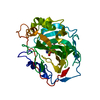
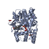
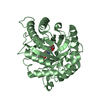
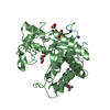
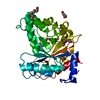
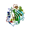
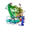
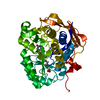
| Title | The structure of GH113 beta-mannanase AaManA from Alicyclobacillus acidocaldarius in complex with ManIFG | ||||||
 Components Components | ENDO-BETA-1,4-MANNANASE | ||||||
 Keywords Keywords | HYDROLASE / BETA-MANNOSIDASE / MANNOSIDASE / GLYCOSIDE HYDROLASE / GH26 / GH113 / CAZY / ENZYME-CARBOHYDRATE INTERACTION / GLYCOSIDASE INHIBITION / QUANTUM MECHANICS / BIOCATALYSIS / CONFORMATION | ||||||
| Function / homology |  Function and homology information Function and homology information: / Glycoside Hydrolase Family 113 / Glycosidases / Glycoside hydrolase superfamily / TIM Barrel / Alpha-Beta Barrel / Alpha Beta Similarity search - Domain/homology | ||||||
| Biological species |  ALICYCLOBACILLUS ACIDOCALDARIUS (bacteria) ALICYCLOBACILLUS ACIDOCALDARIUS (bacteria) | ||||||
| Method |  X-RAY DIFFRACTION / SYNCHROTRON / MOLECULAR REPLACEMENT / Resolution: 1.64 Å X-RAY DIFFRACTION / SYNCHROTRON / MOLECULAR REPLACEMENT / Resolution: 1.64 Å | ||||||
 Authors Authors | Williams, R.J. / Iglesias-Fernandez, J. / Stepper, J. / Jackson, A. / Thompson, A.J. / Lowe, E.C. / White, J.M. / Gilbert, H.J. / Rovira, C. / Davies, G.J. / Williams, S.J. | ||||||
 Citation Citation | Journal: Angew.Chem.Int.Ed.Engl. / Year: 2014 Title: Combined Inhibitor Free-Energy Landscape and Structural Analysis Reports on the Mannosidase Conformational Coordinate. Authors: Williams, R.J. / Iglesias-Fernandez, J. / Stepper, J. / Jackson, A. / Thompson, A.J. / Lowe, E.C. / White, J.M. / Gilbert, H.J. / Rovira, C. / Davies, G.J. / Williams, S.J. | ||||||
| History |
| ||||||
| Remark 700 | SHEET DETERMINATION METHOD: DSSP THE SHEETS PRESENTED AS "AA" IN EACH CHAIN ON SHEET RECORDS BELOW ... SHEET DETERMINATION METHOD: DSSP THE SHEETS PRESENTED AS "AA" IN EACH CHAIN ON SHEET RECORDS BELOW IS ACTUALLY AN 8-STRANDED BARREL THIS IS REPRESENTED BY A 9-STRANDED SHEET IN WHICH THE FIRST AND LAST STRANDS ARE IDENTICAL. |
- Structure visualization
| Structure viewer | Molecule: MolmilJmol/JSmol |
|---|
- Downloads & links
-Download
| PDBx/mmCIF format | 4cd6.cif.gz | 147.1 KB | Display | PDBx/mmCIF format |
|---|---|---|---|---|
| PDB format | pdb4cd6.ent.gz | 114.6 KB | Display | PDB format |
| PDBx/mmJSON format | 4cd6.json.gz | Tree view | PDBx/mmJSON format | |
| Others |  Other downloads Other downloads |
-Validation report
| Arichive directory | https://data.pdbj.org/pub/pdb/validation_reports/cd/4cd6ftp://data.pdbj.org/pub/pdb/validation_reports/cd/4cd6 | HTTPS FTP |
|---|
-Related structure data
| Related structure data | 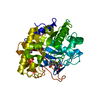 4cd4C 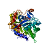 4cd5C 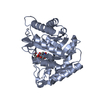 4cd7C  4cd8C 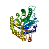 3civS C: citing same article ( S: Starting model for refinement |
|---|---|
| Similar structure data |


-Links
 PDBj
PDBj- Assembly
| Deposited unit | 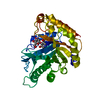
| ||||||||
|---|---|---|---|---|---|---|---|---|---|
| 1 |
| ||||||||
| Unit cell |
|
-Components
| #1: Protein | Mass: 36342.961 Da / Num. of mol.: 1 Source method: isolated from a genetically manipulated source Source: (gene. exp.) ALICYCLOBACILLUS ACIDOCALDARIUS (bacteria)Strain: TC-12-31 / Production host: References: UniProt: A5H1I6, mannan endo-1,4-beta-mannosidase |
|---|---|
| #2: Chemical | ChemComp-IFM /   Mass: 147.172 Da / Num. of mol.: 1 / Source method: obtained synthetically / Formula: C6H13NO3 Mass: 147.172 Da / Num. of mol.: 1 / Source method: obtained synthetically / Formula: C6H13NO3 |
| #3: Sugar | ChemComp-BMA /   Type: D-saccharide, beta linking / Mass: 180.156 Da / Num. of mol.: 1 Type: D-saccharide, beta linking / Mass: 180.156 Da / Num. of mol.: 1Source method: isolated from a genetically manipulated source Formula: C6H12O6 |
| #4: Water | ChemComp-HOH /  Mass: 18.015 Da / Num. of mol.: 189 / Source method: isolated from a natural source / Formula: H2O Mass: 18.015 Da / Num. of mol.: 189 / Source method: isolated from a natural source / Formula: H2O |
-Experimental details
-Experiment
| Experiment | Method: X-RAY DIFFRACTION / Number of used crystals: 1 |
|---|
- Sample preparation
| Crystal | Density Matthews: 2.17 Å3/Da / Density % sol: 43 % / Description: NONE |
|---|---|
| Crystal grow | pH: 4.6 / Details: 0.1 M SODIUM ACETATE PH 4.6, 4% PEG 4000 |
-Data collection
| Diffraction | Mean temperature: 100 K |
|---|---|
| Diffraction source | Source: SYNCHROTRON / Site: Diamond  / Beamline: I03 / Wavelength: 0.9762 / Beamline: I03 / Wavelength: 0.9762 |
| Detector | Type: DECTRIS PILATUS 6M / Detector: PIXEL / Date: Apr 19, 2013 / Details: MIRRORS |
| Radiation | Protocol: SINGLE WAVELENGTH / Monochromatic (M) / Laue (L): M / Scattering type: x-ray |
| Radiation wavelength | Wavelength: 0.9762 Å / Relative weight: 1 |
| Reflection | Resolution: 1.64→46.87 Å / Num. obs: 39738 / % possible obs: 99.4 % / Observed criterion σ(I): 2 / Redundancy: 6.3 % / Rmerge(I) obs: 0.07 / Net I/σ(I): 12.7 |
| Reflection shell | Resolution: 1.64→1.69 Å / Redundancy: 5.7 % / Rmerge(I) obs: 0.61 / Mean I/σ(I) obs: 2.5 / % possible all: 99.1 |
- Processing
| Software |
| ||||||||||||||||||||||||||||||||||||||||||||||||||||||||||||||||||||||||||||||||||||||||||||||||||||||||||||||||||||||||||||||||||||||||||||||||||||||||||||||||||||||||||||||||||||||
|---|---|---|---|---|---|---|---|---|---|---|---|---|---|---|---|---|---|---|---|---|---|---|---|---|---|---|---|---|---|---|---|---|---|---|---|---|---|---|---|---|---|---|---|---|---|---|---|---|---|---|---|---|---|---|---|---|---|---|---|---|---|---|---|---|---|---|---|---|---|---|---|---|---|---|---|---|---|---|---|---|---|---|---|---|---|---|---|---|---|---|---|---|---|---|---|---|---|---|---|---|---|---|---|---|---|---|---|---|---|---|---|---|---|---|---|---|---|---|---|---|---|---|---|---|---|---|---|---|---|---|---|---|---|---|---|---|---|---|---|---|---|---|---|---|---|---|---|---|---|---|---|---|---|---|---|---|---|---|---|---|---|---|---|---|---|---|---|---|---|---|---|---|---|---|---|---|---|---|---|---|---|---|---|
| Refinement | Method to determine structure: MOLECULAR REPLACEMENT Starting model: PDB ENTRY 3CIV Resolution: 1.64→46.87 Å / Cor.coef. Fo:Fc: 0.979 / Cor.coef. Fo:Fc free: 0.959 / SU B: 5.624 / SU ML: 0.08 / Cross valid method: THROUGHOUT / ESU R: 0.107 / ESU R Free: 0.093 / Stereochemistry target values: MAXIMUM LIKELIHOOD Details: HYDROGENS HAVE BEEN ADDED IN THE RIDING POSITIONS. U VALUES REFINED INDIVIDUALLY.
| ||||||||||||||||||||||||||||||||||||||||||||||||||||||||||||||||||||||||||||||||||||||||||||||||||||||||||||||||||||||||||||||||||||||||||||||||||||||||||||||||||||||||||||||||||||||
| Solvent computation | Ion probe radii: 0.8 Å / Shrinkage radii: 0.8 Å / VDW probe radii: 1.2 Å / Solvent model: MASK | ||||||||||||||||||||||||||||||||||||||||||||||||||||||||||||||||||||||||||||||||||||||||||||||||||||||||||||||||||||||||||||||||||||||||||||||||||||||||||||||||||||||||||||||||||||||
| Displacement parameters | Biso mean: 32.018 Å2
| ||||||||||||||||||||||||||||||||||||||||||||||||||||||||||||||||||||||||||||||||||||||||||||||||||||||||||||||||||||||||||||||||||||||||||||||||||||||||||||||||||||||||||||||||||||||
| Refinement step | Cycle: LAST / Resolution: 1.64→46.87 Å
| ||||||||||||||||||||||||||||||||||||||||||||||||||||||||||||||||||||||||||||||||||||||||||||||||||||||||||||||||||||||||||||||||||||||||||||||||||||||||||||||||||||||||||||||||||||||
| Refine LS restraints |
|