 Movie
Movie Controller


+ Open data

- Basic information
| Entry | Database: PDB / ID: 4c21 | ||||||
|---|---|---|---|---|---|---|---|
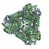
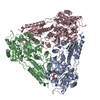
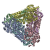
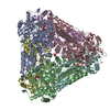
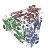
| Title | L-Fucose Isomerase In Complex With Fucitol | ||||||
 Components Components | L-FUCOSE ISOMERASE | ||||||
 Keywords Keywords | ISOMERASE / FUCOSE PROCESSING | ||||||
| Function / homology |  Function and homology information Function and homology informationL-fucose isomerase / L-fucose isomerase activity / arabinose isomerase activity / D-arabinose catabolic process / L-fucose catabolic process / manganese ion binding / cytoplasm Similarity search - Function | ||||||
| Biological species |   STREPTOCOCCUS PNEUMONIAE (bacteria) STREPTOCOCCUS PNEUMONIAE (bacteria) | ||||||
| Method |  X-RAY DIFFRACTION / SYNCHROTRON / MOLECULAR REPLACEMENT / Resolution: 2.55 Å X-RAY DIFFRACTION / SYNCHROTRON / MOLECULAR REPLACEMENT / Resolution: 2.55 Å | ||||||
 Authors Authors | Higgins, M.A. / Suits, M.D.L. / Marsters, C. / Boraston, A.B. | ||||||
 Citation Citation | Journal: J.Mol.Biol. / Year: 2014 Title: Structural and Functional Analysis of Fucose-Processing Enzymes from Streptococcus Pneumoniae. Authors: Higgins, M.A. / Suits, M.D. / Marsters, C. / Boraston, A.B. | ||||||
| History |
| ||||||
| Remark 700 | SHEET DETERMINATION METHOD: DSSP THE SHEETS PRESENTED AS "AD" IN EACH CHAIN ON SHEET RECORDS BELOW ... SHEET DETERMINATION METHOD: DSSP THE SHEETS PRESENTED AS "AD" IN EACH CHAIN ON SHEET RECORDS BELOW IS ACTUALLY AN 6-STRANDED BARREL THIS IS REPRESENTED BY A 7-STRANDED SHEET IN WHICH THE FIRST AND LAST STRANDS ARE IDENTICAL. THE SHEETS PRESENTED AS "BE" IN EACH CHAIN ON SHEET RECORDS BELOW IS ACTUALLY AN 6-STRANDED BARREL THIS IS REPRESENTED BY A 7-STRANDED SHEET IN WHICH THE FIRST AND LAST STRANDS ARE IDENTICAL. |
- Structure visualization
| Structure viewer | Molecule: MolmilJmol/JSmol |
|---|
- Downloads & links
-Download
| PDBx/mmCIF format | 4c21.cif.gz | 260.3 KB | Display | PDBx/mmCIF format |
|---|---|---|---|---|
| PDB format | pdb4c21.ent.gz | 207 KB | Display | PDB format |
| PDBx/mmJSON format | 4c21.json.gz | Tree view | PDBx/mmJSON format | |
| Others |  Other downloads Other downloads |
-Validation report
| Arichive directory | https://data.pdbj.org/pub/pdb/validation_reports/c2/4c21ftp://data.pdbj.org/pub/pdb/validation_reports/c2/4c21 | HTTPS FTP |
|---|
-Related structure data
| Related structure data |  4c20C  4c22C  4c23C  4c24C  4c25C  1fuiS C: citing same article ( S: Starting model for refinement |
|---|---|
| Similar structure data |


-Links
 PDBj
PDBj- Assembly
| Deposited unit | 
| ||||||||||||
|---|---|---|---|---|---|---|---|---|---|---|---|---|---|
| 1 | 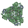
| ||||||||||||
| Unit cell |
| ||||||||||||
| Components on special symmetry positions |
|
-Components
| #1: Protein | Mass: 67825.578 Da / Num. of mol.: 2 Source method: isolated from a genetically manipulated source Source: (gene. exp.) STREPTOCOCCUS PNEUMONIAE (bacteria) / Strain: TIGR4 / Plasmid: PET28 / Production host: #2: Chemical |   Mass: 54.938 Da / Num. of mol.: 2 / Source method: obtained synthetically / Formula: Mn Mass: 54.938 Da / Num. of mol.: 2 / Source method: obtained synthetically / Formula: Mn#3: Chemical |   Mass: 166.172 Da / Num. of mol.: 2 / Source method: obtained synthetically / Formula: C6H14O5 Mass: 166.172 Da / Num. of mol.: 2 / Source method: obtained synthetically / Formula: C6H14O5#4: Chemical | ChemComp-EDO /   Mass: 62.068 Da / Num. of mol.: 16 / Source method: obtained synthetically / Formula: C2H6O2 Mass: 62.068 Da / Num. of mol.: 16 / Source method: obtained synthetically / Formula: C2H6O2#5: Water | ChemComp-HOH / |  Mass: 18.015 Da / Num. of mol.: 680 / Source method: isolated from a natural source / Formula: H2O Mass: 18.015 Da / Num. of mol.: 680 / Source method: isolated from a natural source / Formula: H2OSequence details | MOLECULE B INCLUDES THE N-TERMINAL TAG | |
|---|
-Experimental details
-Experiment
| Experiment | Method: X-RAY DIFFRACTION / Number of used crystals: 1 |
|---|
- Sample preparation
| Crystal | Density Matthews: 3.44 Å3/Da / Density % sol: 64.27 % / Description: NONE |
|---|---|
| Crystal grow | pH: 8 Details: SPFCSI AT A CONCENTRATION OF 12 MG ML-1, BUFFERED IN 20 MM TRIS-HCL (PH 7.0), 150 MM NACL, AND 2 MM DTT WAS CRYSTALLIZED BY MIXING WITH EQUAL VOLUMES OF 100 MM TRIS-HCL (PH 8.0), 27% (W PER ...Details: SPFCSI AT A CONCENTRATION OF 12 MG ML-1, BUFFERED IN 20 MM TRIS-HCL (PH 7.0), 150 MM NACL, AND 2 MM DTT WAS CRYSTALLIZED BY MIXING WITH EQUAL VOLUMES OF 100 MM TRIS-HCL (PH 8.0), 27% (W PER V) POLYETHYLENE GLYCOL 4,000, 50 MM NACL, AND 200 MM MAGNESIUM SULPHATE |
-Data collection
| Diffraction | Mean temperature: 100 K |
|---|---|
| Diffraction source | Source: SYNCHROTRON / Site: CLSI  / Beamline: 08ID-1 / Wavelength: 1.03318 / Beamline: 08ID-1 / Wavelength: 1.03318 |
| Detector | Type: MARRESEARCH MX-300HE / Detector: CCD / Date: Mar 27, 2012 Details: COLLIMATING MIRROR WITH TWO STRIPES (SI, RH AND PT) , TOROIDAL FOCUSING MIRROR (RH AND PT) |
| Radiation | Monochromator: KOHZU DOUBLE CRYSTAL MONOCHROMATOR (DCM), FEATURING INDIRECTLY WATER- COOLED FIRST CRYSTAL AND FLAT, LONG SECOND CRYSTAL Protocol: SINGLE WAVELENGTH / Monochromatic (M) / Laue (L): M / Scattering type: x-ray |
| Radiation wavelength | Wavelength: 1.03318 Å / Relative weight: 1 |
| Reflection | Resolution: 2.55→50 Å / Num. obs: 59825 / % possible obs: 99.5 % / Observed criterion σ(I): 0 / Redundancy: 9.8 % / Rmerge(I) obs: 0.11 / Net I/σ(I): 17.4 |
| Reflection shell | Resolution: 2.55→2.69 Å / Redundancy: 8.2 % / Rmerge(I) obs: 0.55 / Mean I/σ(I) obs: 4.3 / % possible all: 97.3 |
- Processing
| Software |
| ||||||||||||||||||||||||||||||||||||||||||||||||||||||||||||||||||||||||||||||||||||||||||||||||||||||||||||||||||||||||||||||||||||||||||||||||||||||||||||||||||||||||||||||||||||||
|---|---|---|---|---|---|---|---|---|---|---|---|---|---|---|---|---|---|---|---|---|---|---|---|---|---|---|---|---|---|---|---|---|---|---|---|---|---|---|---|---|---|---|---|---|---|---|---|---|---|---|---|---|---|---|---|---|---|---|---|---|---|---|---|---|---|---|---|---|---|---|---|---|---|---|---|---|---|---|---|---|---|---|---|---|---|---|---|---|---|---|---|---|---|---|---|---|---|---|---|---|---|---|---|---|---|---|---|---|---|---|---|---|---|---|---|---|---|---|---|---|---|---|---|---|---|---|---|---|---|---|---|---|---|---|---|---|---|---|---|---|---|---|---|---|---|---|---|---|---|---|---|---|---|---|---|---|---|---|---|---|---|---|---|---|---|---|---|---|---|---|---|---|---|---|---|---|---|---|---|---|---|---|---|
| Refinement | Method to determine structure: MOLECULAR REPLACEMENT Starting model: PDB ENTRY 1FUI Resolution: 2.55→49.71 Å / Cor.coef. Fo:Fc: 0.97 / Cor.coef. Fo:Fc free: 0.945 / SU B: 6.172 / SU ML: 0.132 / Cross valid method: THROUGHOUT / ESU R: 0.277 / ESU R Free: 0.206 / Stereochemistry target values: MAXIMUM LIKELIHOOD Details: HYDROGENS HAVE BEEN ADDED IN THE RIDING POSITIONS. HYDROGENS HAVE BEEN USED IF PRESENT IN THE INPUT. U VALUES REFINED INDIVIDUALLY
| ||||||||||||||||||||||||||||||||||||||||||||||||||||||||||||||||||||||||||||||||||||||||||||||||||||||||||||||||||||||||||||||||||||||||||||||||||||||||||||||||||||||||||||||||||||||
| Solvent computation | Ion probe radii: 0.8 Å / Shrinkage radii: 0.8 Å / VDW probe radii: 1.2 Å / Solvent model: MASK | ||||||||||||||||||||||||||||||||||||||||||||||||||||||||||||||||||||||||||||||||||||||||||||||||||||||||||||||||||||||||||||||||||||||||||||||||||||||||||||||||||||||||||||||||||||||
| Displacement parameters | Biso mean: 33.132 Å2
| ||||||||||||||||||||||||||||||||||||||||||||||||||||||||||||||||||||||||||||||||||||||||||||||||||||||||||||||||||||||||||||||||||||||||||||||||||||||||||||||||||||||||||||||||||||||
| Refinement step | Cycle: LAST / Resolution: 2.55→49.71 Å
| ||||||||||||||||||||||||||||||||||||||||||||||||||||||||||||||||||||||||||||||||||||||||||||||||||||||||||||||||||||||||||||||||||||||||||||||||||||||||||||||||||||||||||||||||||||||
| Refine LS restraints |
|