 Movie
Movie Controller


+ Open data

- Basic information
| Entry | Database: PDB / ID: 4brw | ||||||
|---|---|---|---|---|---|---|---|
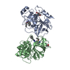
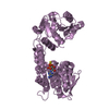
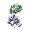
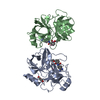
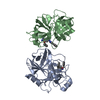
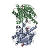
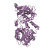
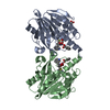
| Title | Crystal structure of the yeast Dhh1-Pat1 complex | ||||||
 Components Components |
| ||||||
 Keywords Keywords | HYDROLASE / TRANSLATIONAL REPRESSION / MRNP REMODELING / P- BOD | ||||||
| Function / homology |  Function and homology information Function and homology informationinvasive filamentous growth / regulation of cytoplasmic mRNA processing body assembly / response to pheromone triggering conjugation with cellular fusion / mRNA decay by 5' to 3' exoribonuclease / pseudohyphal growth / Lsm1-7-Pat1 complex / deadenylation-dependent decapping of nuclear-transcribed mRNA / negative regulation of translational elongation / cytoplasmic side of membrane / filamentous growth ...invasive filamentous growth / regulation of cytoplasmic mRNA processing body assembly / response to pheromone triggering conjugation with cellular fusion / mRNA decay by 5' to 3' exoribonuclease / pseudohyphal growth / Lsm1-7-Pat1 complex / deadenylation-dependent decapping of nuclear-transcribed mRNA / negative regulation of translational elongation / cytoplasmic side of membrane / filamentous growth / formation of translation preinitiation complex / cellular response to nitrogen starvation / P-body assembly / regulation of translational initiation / mRNA transport / cellular response to glucose starvation / negative regulation of translational initiation / stress granule assembly / positive regulation of translation / P-body / kinetochore / mRNA processing / cytoplasmic stress granule / cytosolic small ribosomal subunit / RNA helicase activity / negative regulation of translation / RNA helicase / cell division / mRNA binding / chromatin binding / ATP hydrolysis activity / RNA binding / ATP binding / nucleus / cytoplasm Similarity search - Function | ||||||
| Biological species |  | ||||||
| Method |  X-RAY DIFFRACTION / SYNCHROTRON / MOLECULAR REPLACEMENT / Resolution: 2.795 Å X-RAY DIFFRACTION / SYNCHROTRON / MOLECULAR REPLACEMENT / Resolution: 2.795 Å | ||||||
 Authors Authors | Sharif, H. / Ozgur, S. / Sharma, K. / Basquin, C. / Urlaub, H. / Conti, E. | ||||||
 Citation Citation | Journal: Nucleic Acids Res. / Year: 2013 Title: Structural Analysis of the Yeast Dhh1-Pat1 Complex Reveals How Dhh1 Engages Pat1, Edc3 and RNA in Mutually Exclusive Interactions Authors: Sharif, H. / Ozgur, S. / Sharma, K. / Basquin, C. / Urlaub, H. / Conti, E. | ||||||
| History |
|
- Structure visualization
| Structure viewer | Molecule: MolmilJmol/JSmol |
|---|
- Downloads & links
-Download
| PDBx/mmCIF format | 4brw.cif.gz | 94.6 KB | Display | PDBx/mmCIF format |
|---|---|---|---|---|
| PDB format | pdb4brw.ent.gz | 70.4 KB | Display | PDB format |
| PDBx/mmJSON format | 4brw.json.gz | Tree view | PDBx/mmJSON format | |
| Others |  Other downloads Other downloads |
-Validation report
| Arichive directory | https://data.pdbj.org/pub/pdb/validation_reports/br/4brwftp://data.pdbj.org/pub/pdb/validation_reports/br/4brw | HTTPS FTP |
|---|
-Related structure data
| Related structure data | 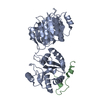 4bruC 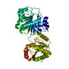 1s2mS C: citing same article ( S: Starting model for refinement |
|---|---|
| Similar structure data |


-Links
 PDBj
PDBj- Assembly
| Deposited unit | 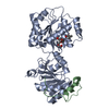
| ||||||||
|---|---|---|---|---|---|---|---|---|---|
| 1 |
| ||||||||
| Unit cell |
|
-Components
| #1: Protein | Mass: 42840.594 Da / Num. of mol.: 1 / Fragment: RESIDUES 46-422 / Mutation: YES Source method: isolated from a genetically manipulated source Source: (gene. exp.) Strain: S288C / Production host:  |
|---|---|
| #2: Protein | Mass: 7912.046 Da / Num. of mol.: 1 / Fragment: N-TERMINAL DOMAIN RESIDUES 5-79 Source method: isolated from a genetically manipulated source Source: (gene. exp.) Strain: S288C / Production host: |
| #3: Chemical | ChemComp-MPD / (  Mass: 118.174 Da / Num. of mol.: 1 / Source method: obtained synthetically / Formula: C6H14O2 / Comment: precipitant*YM Mass: 118.174 Da / Num. of mol.: 1 / Source method: obtained synthetically / Formula: C6H14O2 / Comment: precipitant*YM |
| #4: Chemical | ChemComp-1PE /   Mass: 238.278 Da / Num. of mol.: 1 / Source method: obtained synthetically / Formula: C10H22O6 / Comment: precipitant*YM Mass: 238.278 Da / Num. of mol.: 1 / Source method: obtained synthetically / Formula: C10H22O6 / Comment: precipitant*YM |
| #5: Water | ChemComp-HOH /  Mass: 18.015 Da / Num. of mol.: 4 / Source method: isolated from a natural source / Formula: H2O Mass: 18.015 Da / Num. of mol.: 4 / Source method: isolated from a natural source / Formula: H2O |
-Experimental details
-Experiment
| Experiment | Method: X-RAY DIFFRACTION |
|---|
- Sample preparation
| Crystal | Density Matthews: 3.21 Å3/Da / Density % sol: 61.74 % / Description: NONE |
|---|---|
| Crystal grow | Details: 50 MM TRIS PH 8.0, 4% MPD, 200 MM NACL, 25% PEG 400 |
-Data collection
| Diffraction | Mean temperature: 100 K |
|---|---|
| Diffraction source | Source: SYNCHROTRON / Site: SLS  / Beamline: X10SA / Wavelength: 0.97139 / Beamline: X10SA / Wavelength: 0.97139 |
| Detector | Type: DECTRIS PILATUS 6M / Detector: PIXEL / Date: Feb 25, 2012 |
| Radiation | Protocol: SINGLE WAVELENGTH / Monochromatic (M) / Laue (L): M / Scattering type: x-ray |
| Radiation wavelength | Wavelength: 0.97139 Å / Relative weight: 1 |
| Reflection | Resolution: 2.8→79.88 Å / Num. obs: 17668 / % possible obs: 99.6 % / Redundancy: 8.6 % / Biso Wilson estimate: 73.29 Å2 / Rmerge(I) obs: 0.08 / Net I/σ(I): 18.3 |
| Reflection shell | Resolution: 2.8→2.95 Å / Redundancy: 8.4 % / Rmerge(I) obs: 0.76 / Mean I/σ(I) obs: 2.8 / % possible all: 97.1 |
- Processing
| Software |
| |||||||||||||||||||||||||||||||||||||||||||||||||||||||||||||||||||||||||||||||||||||||||||
|---|---|---|---|---|---|---|---|---|---|---|---|---|---|---|---|---|---|---|---|---|---|---|---|---|---|---|---|---|---|---|---|---|---|---|---|---|---|---|---|---|---|---|---|---|---|---|---|---|---|---|---|---|---|---|---|---|---|---|---|---|---|---|---|---|---|---|---|---|---|---|---|---|---|---|---|---|---|---|---|---|---|---|---|---|---|---|---|---|---|---|---|---|
| Refinement | Method to determine structure: MOLECULAR REPLACEMENT Starting model: PDB ENTRY 1S2M Resolution: 2.795→48.474 Å / SU ML: 0.41 / σ(F): 1.37 / Phase error: 25.94 / Stereochemistry target values: ML / Details: DISORDERED CHAIN B RESIDUES 5-24, 55-79
| |||||||||||||||||||||||||||||||||||||||||||||||||||||||||||||||||||||||||||||||||||||||||||
| Solvent computation | Shrinkage radii: 0.7 Å / VDW probe radii: 1 Å / Solvent model: FLAT BULK SOLVENT MODEL | |||||||||||||||||||||||||||||||||||||||||||||||||||||||||||||||||||||||||||||||||||||||||||
| Displacement parameters | Biso mean: 48.3 Å2 | |||||||||||||||||||||||||||||||||||||||||||||||||||||||||||||||||||||||||||||||||||||||||||
| Refinement step | Cycle: LAST / Resolution: 2.795→48.474 Å
| |||||||||||||||||||||||||||||||||||||||||||||||||||||||||||||||||||||||||||||||||||||||||||
| Refine LS restraints |
| |||||||||||||||||||||||||||||||||||||||||||||||||||||||||||||||||||||||||||||||||||||||||||
| LS refinement shell |
|
