 Movie
Movie Controller

[English] 日本語
 Yorodumi
- PDB-4bkk: The Respiratory Syncytial Virus nucleoprotein-RNA complex forms a... -
+ Open data

- Basic information
| Entry | Database: PDB / ID: 4bkk | ||||||
|---|---|---|---|---|---|---|---|
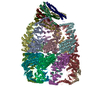
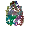
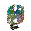
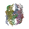
| Title | The Respiratory Syncytial Virus nucleoprotein-RNA complex forms a left-handed helical nucleocapsid. | ||||||
 Components Components |
| ||||||
 Keywords Keywords | VIRAL PROTEIN/RNA / VIRAL PROTEIN-RNA COMPLEX | ||||||
| Function / homology |  Function and homology information Function and homology informationRespiratory syncytial virus genome transcription / Translation of respiratory syncytial virus mRNAs / symbiont-mediated suppression of host PKR/eIFalpha signaling / helical viral capsid / Respiratory syncytial virus genome replication / RSV-host interactions / Assembly and release of respiratory syncytial virus (RSV) virions / Maturation of hRSV A proteins / Respiratory syncytial virus (RSV) attachment and entry / protein serine/threonine kinase inhibitor activity ...Respiratory syncytial virus genome transcription / Translation of respiratory syncytial virus mRNAs / symbiont-mediated suppression of host PKR/eIFalpha signaling / helical viral capsid / Respiratory syncytial virus genome replication / RSV-host interactions / Assembly and release of respiratory syncytial virus (RSV) virions / Maturation of hRSV A proteins / Respiratory syncytial virus (RSV) attachment and entry / protein serine/threonine kinase inhibitor activity / symbiont-mediated suppression of host cytoplasmic pattern recognition receptor signaling pathway via inhibition of MDA-5 activity / symbiont-mediated suppression of host cytoplasmic pattern recognition receptor signaling pathway via inhibition of MAVS activity / PKR-mediated signaling / Evasion by RSV of host interferon responses / viral capsid / viral nucleocapsid / symbiont-mediated suppression of host NF-kappaB cascade / host cell cytoplasm / symbiont-mediated suppression of host type I interferon-mediated signaling pathway / ribonucleoprotein complex / RNA binding Similarity search - Function | ||||||
| Biological species |  HUMAN RESPIRATORY SYNCYTIAL VIRUS A STRAIN LONG HUMAN RESPIRATORY SYNCYTIAL VIRUS A STRAIN LONG | ||||||
| Method | ELECTRON MICROSCOPY / electron tomography / negative staining | ||||||
 Authors Authors | Bakker, S.E. / Duquerroy, S. / Galloux, M. / Loney, C. / Conner, E. / Eleouet, J.F. / Rey, F.A. / Bhella, D. | ||||||
 Citation Citation | Journal: J Gen Virol / Year: 2013 Title: The respiratory syncytial virus nucleoprotein-RNA complex forms a left-handed helical nucleocapsid. Authors: Saskia E Bakker / Stéphane Duquerroy / Marie Galloux / Colin Loney / Edward Conner / Jean-François Eléouët / Félix A Rey / David Bhella /   Abstract: Respiratory syncytial virus (RSV) is an important human pathogen. Its nucleocapsid (NC), which comprises the negative sense RNA viral genome coated by the viral nucleoprotein N, is a critical ...Respiratory syncytial virus (RSV) is an important human pathogen. Its nucleocapsid (NC), which comprises the negative sense RNA viral genome coated by the viral nucleoprotein N, is a critical assembly that serves as template for both mRNA synthesis and genome replication. We have previously described the X-ray structure of an NC-like structure: a decameric ring formed of N-RNA that mimics one turn of the helical NC. In the absence of experimental data we had hypothesized that the NC helix would be right-handed, as the N-N contacts in the ring appeared to more easily adapt to that conformation. We now unambiguously show that the RSV NC is a left-handed helix. We further show that the contacts in the ring can be distorted to maintain key N-N-protein interactions in a left-handed helix, and discuss the implications of the resulting atomic model of the helical NC for viral replication and transcription. | ||||||
| History |
|
- Structure visualization
| Movie |
Movie viewer |
|---|---|
| Structure viewer | Molecule: MolmilJmol/JSmol |
- Downloads & links
-Download
| PDBx/mmCIF format | 4bkk.cif.gz | 1.6 MB | Display | PDBx/mmCIF format |
|---|---|---|---|---|
| PDB format | pdb4bkk.ent.gz | 1.3 MB | Display | PDB format |
| PDBx/mmJSON format | 4bkk.json.gz | Tree view | PDBx/mmJSON format | |
| Others |  Other downloads Other downloads |
-Validation report
| Arichive directory | https://data.pdbj.org/pub/pdb/validation_reports/bk/4bkkftp://data.pdbj.org/pub/pdb/validation_reports/bk/4bkk | HTTPS FTP |
|---|
-Related structure data
| Related structure data |  2369MC  2370C M: map data used to model this data C: citing same article ( |
|---|---|
| Similar structure data |


-Links
 PDBj
PDBj


- Assembly
| Deposited unit | 
|
|---|---|
| 1 |
|
-Components
| #1: RNA chain | Mass: 49089.227 Da / Num. of mol.: 1 Source method: isolated from a genetically manipulated source Source: (gene. exp.) HUMAN RESPIRATORY SYNCYTIAL VIRUS A STRAIN LONGCell line (production host): Sf21 / Production host:   SPODOPTERA FRUGIPERDA (fall armyworm) SPODOPTERA FRUGIPERDA (fall armyworm) |
|---|---|
| #2: Protein | Mass: 43507.848 Da / Num. of mol.: 23 Source method: isolated from a genetically manipulated source Source: (gene. exp.) HUMAN RESPIRATORY SYNCYTIAL VIRUS A STRAIN LONGCell line (production host): Sf21 / Production host: SPODOPTERA FRUGIPERDA (fall armyworm) / References: UniProt: P03418 |
-Experimental details
-Experiment
| Experiment | Method: ELECTRON MICROSCOPY |
|---|---|
| EM experiment | Aggregation state: FILAMENT / 3D reconstruction method: electron tomography |
- Sample preparation
| Component | Name: PURIFIED RECOMBINANT RSV NUCLEOCAPSIDS. / Type: VIRUS |
|---|---|
| Buffer solution | Name: 50 MM TRIS-HCL, PH 7.4, 150 MM NACL, 10 MM EDTA / pH: 7.4 / Details: 50 MM TRIS-HCL, PH 7.4, 150 MM NACL, 10 MM EDTA |
| Specimen | Embedding applied: NO / Shadowing applied: NO / Staining applied: YES / Vitrification applied: NO |
| EM staining | Type: NEGATIVE / Material: Ammonium Molybdate |
| Specimen support | Details: HOLEY CARBON |
- Electron microscopy imaging
| Microscopy | Model: JEOL 2200FSC / Date: Jul 15, 2010 Details: ROOM TEMPERATURE STAINED GRIDS IMAGED AT LN2 TEMPERATURES. |
|---|---|
| Electron gun | Electron source:  FIELD EMISSION GUN / Accelerating voltage: 200 kV / Illumination mode: FLOOD BEAM FIELD EMISSION GUN / Accelerating voltage: 200 kV / Illumination mode: FLOOD BEAM |
| Electron lens | Mode: BRIGHT FIELD / Nominal magnification: 40000 X / Nominal defocus max: 4000 nm / Nominal defocus min: 2000 nm / Cs: 2 mm |
| Specimen holder | Tilt angle max: 60 ° / Tilt angle min: -60 ° |
| Image recording | Electron dose: 131 e/Å2 / Film or detector model: GATAN ULTRASCAN 4000 (4k x 4k) |
| Image scans | Num. digital images: 9 |
| Radiation wavelength | Relative weight: 1 |
- Processing
| EM software |
| ||||||||||||
|---|---|---|---|---|---|---|---|---|---|---|---|---|---|
| 3D reconstruction | Method: SUBTOMOGRAM AVERAGING / Num. of particles: 911 / Nominal pixel size: 6.5 Å Details: SUBTOMOGRAM AVERAGING. NOTE THE COORDINATES ARE BASED ON MODELLING. SUBMISSION BASED ON EXPERIMENTAL DATA FROM EMDB EMD-2369. (DEPOSITION ID: 11640). Symmetry type: HELICAL | ||||||||||||
| Atomic model building | Protocol: OTHER / Details: METHOD--MODELLING REFINEMENT PROTOCOL--MODEL | ||||||||||||
| Atomic model building | PDB-ID: 2WJ8 Accession code: 2WJ8 / Source name: PDB / Type: experimental model | ||||||||||||
| Refinement step | Cycle: LAST
|

