[English] 日本語
Yorodumi
- PDB-4bke: Recombinant human serum albumin with palmitic acid. Synthetic cat... -

+
Open data


ID or keywords:

Loading...

-
Basic information

Entry
Database: PDB / ID: 4bke
Title
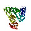
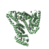
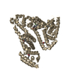
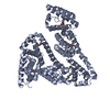
Recombinant human serum albumin with palmitic acid. Synthetic cationic antimicrobial peptides bind with their hydrophobic parts to drug site II of human serum albumin
ComponentsSERUM ALBUMIN
KeywordsTRANSPORT PROTEIN / ALBUMIN BINDING / GROUP EPITOPE MAPPING / MOLECULAR DOCKING
Function / homology
Function and homology information


cellular response to calcium ion starvation / exogenous protein binding / Ciprofloxacin ADME / HDL remodeling / enterobactin binding / Heme biosynthesis / negative regulation of mitochondrial depolarization / Prednisone ADME / Heme degradation / antioxidant activity ...cellular response to calcium ion starvation / exogenous protein binding / Ciprofloxacin ADME / HDL remodeling / enterobactin binding / Heme biosynthesis / negative regulation of mitochondrial depolarization / Prednisone ADME / Heme degradation / antioxidant activity / Aspirin ADME / toxic substance binding / Scavenging of heme from plasma / Recycling of bile acids and salts / cellular response to starvation / platelet alpha granule lumen / fatty acid binding / Post-translational protein phosphorylation / Cytoprotection by HMOX1 / Regulation of Insulin-like Growth Factor (IGF) transport and uptake by Insulin-like Growth Factor Binding Proteins (IGFBPs) / pyridoxal phosphate binding / Platelet degranulation / protein-folding chaperone binding / blood microparticle / copper ion binding / endoplasmic reticulum lumen / Golgi apparatus / endoplasmic reticulum / protein-containing complex / DNA binding / extracellular space / extracellular exosome / extracellular region / identical protein binding / nucleus / cytoplasm
Similarity search - Function
Serum Albumin; Chain A, Domain 1 - #10 / Serum albumin/Alpha-fetoprotein/Afamin / ALB/AFP/VDB / Serum albumin, N-terminal / Serum albumin, conserved site / Serum albumin-like / Serum albumin family / Albumin domain signature. / Albumin domain profile. / serum albumin ...Serum Albumin; Chain A, Domain 1 - #10 / Serum albumin/Alpha-fetoprotein/Afamin / ALB/AFP/VDB / Serum albumin, N-terminal / Serum albumin, conserved site / Serum albumin-like / Serum albumin family / Albumin domain signature. / Albumin domain profile. / serum albumin / Serum Albumin; Chain A, Domain 1 / Orthogonal Bundle / Mainly Alpha
Similarity search - Domain/homology
PALMITIC ACID / Albumin
Similarity search - Component
Biological speciesHOMO SAPIENS (human)
MethodX-RAY DIFFRACTION / MOLECULAR REPLACEMENT / Resolution: 2.35 Å
AuthorsSivertsen, A. / Isaksson, J. / Leiros, H.-K.S. / Svenson, J. / Svendsen, J.-S. / Brandsdal, B.-O.
CitationJournal: Bmc Struct.Biol. / Year: 2014
Title: Synthetic Cationic Antimicrobial Peptides Bind with Their Hydrophobic Parts to Drug Site II of Human Serum Albumin.
Authors: Sivertsen, A. / Isaksson, J. / Leiros, H.S. / Svenson, J. / Svendsen, J. / Brandsdal, B.O.
History
DepositionApr 24, 2013Deposition site: PDBE / Processing site: PDBE
Revision 1.0Feb 5, 2014Provider: repository / Type: Initial release
Revision 1.1Apr 4, 2018Group: Advisory / Data collection / Category: diffrn_source / pdbx_unobs_or_zero_occ_atoms / Item: _diffrn_source.type
Revision 1.2Dec 20, 2023Group: Advisory / Data collection ...Advisory / Data collection / Database references / Derived calculations / Other / Refinement description
Category: chem_comp_atom / chem_comp_bond ...chem_comp_atom / chem_comp_bond / database_2 / pdbx_database_status / pdbx_initial_refinement_model / pdbx_unobs_or_zero_occ_atoms / struct_site
Item: _database_2.pdbx_DOI / _database_2.pdbx_database_accession ..._database_2.pdbx_DOI / _database_2.pdbx_database_accession / _pdbx_database_status.status_code_sf / _struct_site.pdbx_auth_asym_id / _struct_site.pdbx_auth_comp_id / _struct_site.pdbx_auth_seq_id

-
Structure visualization

Structure viewerMolecule:
MolmilJmol/JSmol

Downloads & links

-
Assembly

Deposited unit
A: SERUM ALBUMIN
hetero molecules


Theoretical massNumber of molelcules
Total (without water)71,2658
Polymers69,4701
Non-polymers1,7957
Water46826
1


  • Idetical with deposited unit
  • defined by author&software
TypeNameSymmetry operationNumber
identity operation1_555x,y,z1
MethodPISA
Unit cell
Length a, b, c (Å)189.366, 39.003, 96.296
Angle α, β, γ (deg.)90.00, 105.48, 90.00
Int Tables number5
Space group name H-MC121

-
Components

#1: Protein SERUM ALBUMIN /


Mass: 69469.695 Da / Num. of mol.: 1
Source method: isolated from a genetically manipulated source
Source: (gene. exp.) HOMO SAPIENS (human) / Tissue: BLOOD / Production host: SACCHAROMYCES CEREVISIAE (brewer's yeast) / References: UniProt: P02768
#2: Chemical
ChemComp-PLM / PALMITIC ACID / Palmitic acid


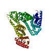
Mass: 256.424 Da / Num. of mol.: 7 / Source method: obtained synthetically / Formula: C16H32O2
#3: Water ChemComp-HOH / water / Water


Mass: 18.015 Da / Num. of mol.: 26 / Source method: isolated from a natural source / Formula: H2O

-
Experimental details

-
Experiment

ExperimentMethod: X-RAY DIFFRACTION / Number of used crystals: 1

-
Sample preparation

CrystalDensity Matthews: 2.47 Å3/Da / Density % sol: 50.13 % / Description: NONE
Crystal growpH: 7.2
Details: 26 % PEG 3350, 50 MM KH2PO4 PH 7.2; 120 MG/ML IN 20 MM KH2PO4 AT PH 7.5 CONTAINING NOMINAL 0.1 MM PALMITIC ACID STREAK SEEDED SEEDING

-
Data collection

DiffractionMean temperature: 295 K
Diffraction sourceSource: ROTATING ANODE / Type: RIGAKU / Wavelength: 1.5418
DetectorType: RIGAKU R-AXIS IV / Detector: IMAGE PLATE
RadiationProtocol: SINGLE WAVELENGTH / Monochromatic (M) / Laue (L): M / Scattering type: x-ray
Radiation wavelengthWavelength: 1.5418 Å / Relative weight: 1
ReflectionResolution: 2.35→38.14 Å / Num. obs: 27926 / % possible obs: 96.5 % / Observed criterion σ(I): 0 / Redundancy: 5 % / Rmerge(I) obs: 0.07 / Net I/σ(I): 9.3
Reflection shellResolution: 2.35→2.48 Å / Redundancy: 5 % / Rmerge(I) obs: 0.66 / Mean I/σ(I) obs: 2.2 / % possible all: 95.2

-
Processing

Software
NameVersionClassification
REFMAC5.5.0102refinement
XDSdata reduction
SCALAdata scaling
RefinementMethod to determine structure: MOLECULAR REPLACEMENT
Starting model: PDB ENTRY 1E7H
Resolution: 2.35→20 Å / Cor.coef. Fo:Fc: 0.962 / Cor.coef. Fo:Fc free: 0.948 / SU B: 8.942 / SU ML: 0.211 / Cross valid method: THROUGHOUT / ESU R: 0.379 / ESU R Free: 0.262 / Stereochemistry target values: MAXIMUM LIKELIHOOD / Details: HYDROGENS HAVE BEEN ADDED IN THE RIDING POSITIONS.
RfactorNum. reflection% reflectionSelection details
Rfree0.25434 2006 7.2 %RANDOM
Rwork0.1975 ---
obs0.20167 25867 96.63 %-
Solvent computationIon probe radii: 0.8 Å / Shrinkage radii: 0.8 Å / VDW probe radii: 1.4 Å / Solvent model: MASK
Displacement parametersBiso mean: 56.95 Å2
Baniso -1Baniso -2Baniso -3
1-2.6 Å20 Å20.34 Å2
2---0.64 Å20 Å2
3----1.78 Å2
Refinement stepCycle: LAST / Resolution: 2.35→20 Å
ProteinNucleic acidLigandSolventTotal
Num. atoms4630 0 119 26 4775
Refine LS restraints
Refine-IDTypeDev idealDev ideal targetNumber
X-RAY DIFFRACTIONr_bond_refined_d0.0090.0224683
X-RAY DIFFRACTIONr_bond_other_d
X-RAY DIFFRACTIONr_angle_refined_deg1.1251.9816321
X-RAY DIFFRACTIONr_angle_other_deg
X-RAY DIFFRACTIONr_dihedral_angle_1_deg4.7645585
X-RAY DIFFRACTIONr_dihedral_angle_2_deg33.75824.505202
X-RAY DIFFRACTIONr_dihedral_angle_3_deg17.99715753
X-RAY DIFFRACTIONr_dihedral_angle_4_deg19.3021523
X-RAY DIFFRACTIONr_chiral_restr0.0790.2699
X-RAY DIFFRACTIONr_gen_planes_refined0.0040.0213496
X-RAY DIFFRACTIONr_gen_planes_other
X-RAY DIFFRACTIONr_nbd_refined
X-RAY DIFFRACTIONr_nbd_other
X-RAY DIFFRACTIONr_nbtor_refined
X-RAY DIFFRACTIONr_nbtor_other
X-RAY DIFFRACTIONr_xyhbond_nbd_refined
X-RAY DIFFRACTIONr_xyhbond_nbd_other
X-RAY DIFFRACTIONr_metal_ion_refined
X-RAY DIFFRACTIONr_metal_ion_other
X-RAY DIFFRACTIONr_symmetry_vdw_refined
X-RAY DIFFRACTIONr_symmetry_vdw_other
X-RAY DIFFRACTIONr_symmetry_hbond_refined
X-RAY DIFFRACTIONr_symmetry_hbond_other
X-RAY DIFFRACTIONr_symmetry_metal_ion_refined
X-RAY DIFFRACTIONr_symmetry_metal_ion_other
X-RAY DIFFRACTIONr_mcbond_it0.5351.52925
X-RAY DIFFRACTIONr_mcbond_other
X-RAY DIFFRACTIONr_mcangle_it1.03524685
X-RAY DIFFRACTIONr_mcangle_other
X-RAY DIFFRACTIONr_scbond_it1.4331758
X-RAY DIFFRACTIONr_scbond_other
X-RAY DIFFRACTIONr_scangle_it2.4964.51636
X-RAY DIFFRACTIONr_scangle_other
X-RAY DIFFRACTIONr_long_range_B_refined
X-RAY DIFFRACTIONr_long_range_B_other
X-RAY DIFFRACTIONr_rigid_bond_restr
X-RAY DIFFRACTIONr_sphericity_free
X-RAY DIFFRACTIONr_sphericity_bonded
LS refinement shellResolution: 2.35→2.41 Å / Total num. of bins used: 20
RfactorNum. reflection% reflection
Rfree0.323 166 -
Rwork0.289 1804 -
obs--93.99 %

+
About Yorodumi

-
News

-
Feb 9, 2022. New format data for meta-information of EMDB entries

New format data for meta-information of EMDB entries

  • Version 3 of the EMDB header file is now the official format.
  • The previous official version 1.9 will be removed from the archive.

Related info.:EMDB header

External links:wwPDB to switch to version 3 of the EMDB data model

-
Aug 12, 2020. Covid-19 info

Covid-19 info

URL: https://pdbj.org/emnavi/covid19.php

New page: Covid-19 featured information page in EM Navigator.

Related info.:Covid-19 info / Mar 5, 2020. Novel coronavirus structure data

+
Mar 5, 2020. Novel coronavirus structure data

Novel coronavirus structure data

Related info.:Yorodumi Speices / Aug 12, 2020. Covid-19 info

External links:COVID-19 featured content - PDBj / Molecule of the Month (242):Coronavirus Proteases

+
Jan 31, 2019. EMDB accession codes are about to change! (news from PDBe EMDB page)

EMDB accession codes are about to change! (news from PDBe EMDB page)

  • The allocation of 4 digits for EMDB accession codes will soon come to an end. Whilst these codes will remain in use, new EMDB accession codes will include an additional digit and will expand incrementally as the available range of codes is exhausted. The current 4-digit format prefixed with “EMD-” (i.e. EMD-XXXX) will advance to a 5-digit format (i.e. EMD-XXXXX), and so on. It is currently estimated that the 4-digit codes will be depleted around Spring 2019, at which point the 5-digit format will come into force.
  • The EM Navigator/Yorodumi systems omit the EMD- prefix.

Related info.:Q: What is EMD? / ID/Accession-code notation in Yorodumi/EM Navigator

External links:EMDB Accession Codes are Changing Soon! / Contact to PDBj

+
Jul 12, 2017. Major update of PDB

Major update of PDB

  • wwPDB released updated PDB data conforming to the new PDBx/mmCIF dictionary.
  • This is a major update changing the version number from 4 to 5, and with Remediation, in which all the entries are updated.
  • In this update, many items about electron microscopy experimental information are reorganized (e.g. em_software).
  • Now, EM Navigator and Yorodumi are based on the updated data.

External links:wwPDB Remediation / Enriched Model Files Conforming to OneDep Data Standards Now Available in the PDB FTP Archive

-
Yorodumi

Thousand views of thousand structures

  • Yorodumi is a browser for structure data from EMDB, PDB, SASBDB, etc.
  • This page is also the successor to EM Navigator detail page, and also detail information page/front-end page for Omokage search.
  • The word "yorodu" (or yorozu) is an old Japanese word meaning "ten thousand". "mi" (miru) is to see.

Related info.:EMDB / PDB / SASBDB / Comparison of 3 databanks / Yorodumi Search / Aug 31, 2016. New EM Navigator & Yorodumi / Yorodumi Papers / Jmol/JSmol / Function and homology information / Changes in new EM Navigator and Yorodumi

Read more