- PDB-4bhx: Crystal structure of the SCAN domain from human paternally expres... -
+
Open data
ID or keywords:
Loading...
-
Basic information
Entry
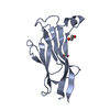
Database: PDB / ID: 4bhx
Title
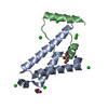
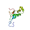
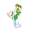
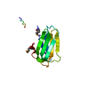
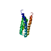
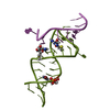
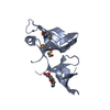
Crystal structure of the SCAN domain from human paternally expressed gene 3 protein
Components
PATERNALLY-EXPRESSED GENE 3 PROTEIN
Keywords
DNA BINDING PROTEIN / PEG3
Function / homology
Function and homology information
autophagosome / RNA polymerase II transcription regulatory region sequence-specific DNA binding / regulation of gene expression / DNA-binding transcription factor activity, RNA polymerase II-specific / apoptotic process / regulation of transcription by RNA polymerase II / negative regulation of transcription by RNA polymerase II / positive regulation of transcription by RNA polymerase II / zinc ion binding / nucleus Similarity search - Function
Mass: 18.015 Da / Num. of mol.: 155 / Source method: isolated from a natural source / Formula: H2O
Sequence details
THE FIRST THREE RESIDUES AT THE N-TERMINUS (GHM) ARE FROM THE HISTIDINE TAG AND REMAINED AFTER TEV DIGESTION.
-
Experimental details
-
Experiment
Experiment
Method: X-RAY DIFFRACTION / Number of used crystals: 1
-
Sample preparation
Crystal
Density Matthews: 2.44 Å3/Da / Density % sol: 49 % / Description: NONE
Crystal grow
Details: CRYSTALLIZATION WAS OBSERVED DURING PROTEIN CONCENTRATION USING VIVASPIN 20 CONCENTRATORS (SARTORIUS STEDIM BIOTECH) IN 50 MM TRIS-HCL PH 7.5, 150 MM NACL.
Resolution: 1.95→13.82 Å / Cor.coef. Fo:Fc: 0.959 / Cor.coef. Fo:Fc free: 0.933 / SU B: 3.489 / SU ML: 0.101 / Cross valid method: THROUGHOUT / ESU R: 0.036 / ESU R Free: 0.033 / Stereochemistry target values: MAXIMUM LIKELIHOOD / Details: HYDROGENS HAVE BEEN ADDED IN THE RIDING POSITIONS.
Rfactor
Num. reflection
% reflection
Selection details
Rfree
0.22383
806
5 %
RANDOM
Rwork
0.17151
-
-
-
obs
0.17412
15239
99.53 %
-
Solvent computation
Ion probe radii: 0.8 Å / Shrinkage radii: 0.8 Å / VDW probe radii: 1.2 Å / Solvent model: MASK
 Movie
Movie Controller

 Yorodumi
 Open data
 Basic information
 Components
 Keywords
 Function and homology information
 HOMO SAPIENS (human)
 X-RAY DIFFRACTION /
  Authors
 Citation
 Structure visualization
 Downloads & links
 Other downloads
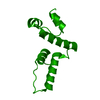


 PDBj
PDBj


 Assembly
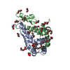


 Mass: 106.120 Da / Num. of mol.: 4 / Source method: obtained synthetically / Formula: C4H10O3
 Mass: 150.173 Da / Num. of mol.: 2 / Source method: obtained synthetically / Formula: C6H14O4
 Mass: 62.068 Da / Num. of mol.: 21 / Source method: obtained synthetically / Formula: C2H6O2
 Mass: 18.015 Da / Num. of mol.: 155 / Source method: isolated from a natural source / Formula: H2O
 Sample preparation
 Processing