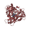
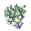
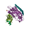
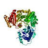
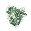
Mass: 69775.000 Da / Num. of mol.: 1 / Mutation: YES Source method: isolated from a genetically manipulated source Source: (gene. exp.) HOMO SAPIENS (human) / Plasmid: PDEST8 / Cell line (production host): High Five / Production host: TRICHOPLUSIA NI (cabbage looper) / References: UniProt: Q14397
Mass: 18.015 Da / Num. of mol.: 700 / Source method: isolated from a natural source / Formula: H2O
Sequence details
SURFACE MUTATION K326T, K327T C-TERMINAL LEHHHHHH TAG
-
Experimental details
-
Experiment
Experiment
Method: X-RAY DIFFRACTION / Number of used crystals: 1
-
Sample preparation
Crystal
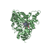
Density Matthews: 2.16 Å3/Da / Density % sol: 43.11 % / Description: NONE
Crystal grow
Details: 12-16 MG/ML IN 25 MM HEPES PH 7.4, 50 MM KCL, 1 MM MGCL2, 2 MM DTT AND 5 MM FRUCTOSE-1-PHOSPHATE 14 % PEG 8000, 20% GLYCEROL, 0.16 M CALCIUM ACETATE AND 0.08 M CACODYLATE PH 6.5
Protocol: SINGLE WAVELENGTH / Monochromatic (M) / Laue (L): M / Scattering type: x-ray
Radiation wavelength
Wavelength: 0.96 Å / Relative weight: 1
Reflection
Resolution: 1.47→72 Å / Num. obs: 102068 / % possible obs: 98.6 % / Observed criterion σ(I): -3 / Redundancy: 3.5 % / Biso Wilson estimate: 15.5 Å2 / Rmerge(I) obs: 0.05 / Net I/σ(I): 14.7
Reflection shell
Resolution: 1.47→1.53 Å / Rmerge(I) obs: 0.4 / Mean I/σ(I) obs: 3.9 / % possible all: 98.2
-
Processing
Software
Name
Version
Classification
BUSTER
2.9.3
refinement
XDS
datareduction
XSCALE
datascaling
autoSHARP
phasing
Refinement
Method to determine structure: SIRAS Starting model: NONE Resolution: 1.47→21.52 Å / Cor.coef. Fo:Fc: 0.9644 / Cor.coef. Fo:Fc free: 0.9581 / SU R Cruickshank DPI: 0.061 / Cross valid method: THROUGHOUT / σ(F): 0 / SU R Blow DPI: 0.065 / SU Rfree Blow DPI: 0.063 / SU Rfree Cruickshank DPI: 0.06 Details: NUMBER OF LIBRARIES USED: 7. IDEAL-DIST CONTACT TERM CONTACT SETUP. RESIDUE TYPES WITHOUT CCP4 ATOM TYPE IN LIBRARY=F1P CA. NUMBER OF ATOMS WITH PROPER CCP4 ATOM TYPE= 5340. NUMBER WITH ...Details: NUMBER OF LIBRARIES USED: 7. IDEAL-DIST CONTACT TERM CONTACT SETUP. RESIDUE TYPES WITHOUT CCP4 ATOM TYPE IN LIBRARY=F1P CA. NUMBER OF ATOMS WITH PROPER CCP4 ATOM TYPE= 5340. NUMBER WITH APPROX DEFAULT CCP4 ATOM TYPE=16. NUMBER TREATED BY BAD NON-BONDED CONTACTS=1.
Rfactor
Num. reflection
% reflection
Selection details
Rfree
0.1774
5102
5 %
RANDOM
Rwork
0.16
-
-
-
obs
0.1609
102029
98.58 %
-
Displacement parameters
Biso mean: 18.61 Å2
Baniso -1
Baniso -2
Baniso -3
1-
0.9777 Å2
0 Å2
0 Å2
2-
-
0.6609 Å2
0 Å2
3-
-
-
-1.6386 Å2
Refine analyze
Luzzati coordinate error obs: 0.145 Å
Refinement step
Cycle: LAST / Resolution: 1.47→21.52 Å
Protein
Nucleic acid
Ligand
Solvent
Total
Num. atoms
4606
0
17
700
5323
Refine LS restraints
Refine-ID
Type
Dev ideal
Number
Restraint function
Weight
X-RAY DIFFRACTION
t_bond_d
0.008
4747
HARMONIC
2
X-RAY DIFFRACTION
t_angle_deg
0.95
6441
HARMONIC
2
X-RAY DIFFRACTION
t_dihedral_angle_d
1670
SINUSOIDAL
2
X-RAY DIFFRACTION
t_incorr_chiral_ct
X-RAY DIFFRACTION
t_pseud_angle
X-RAY DIFFRACTION
t_trig_c_planes
114
HARMONIC
2
X-RAY DIFFRACTION
t_gen_planes
688
HARMONIC
5
X-RAY DIFFRACTION
t_it
4747
HARMONIC
20
X-RAY DIFFRACTION
t_nbd
0
SEMIHARMONIC
5
X-RAY DIFFRACTION
t_omega_torsion
3.35
X-RAY DIFFRACTION
t_other_torsion
14.05
X-RAY DIFFRACTION
t_improper_torsion
X-RAY DIFFRACTION
t_chiral_improper_torsion
644
SEMIHARMONIC
5
X-RAY DIFFRACTION
t_sum_occupancies
X-RAY DIFFRACTION
t_utility_distance
X-RAY DIFFRACTION
t_utility_angle
X-RAY DIFFRACTION
t_utility_torsion
X-RAY DIFFRACTION
t_ideal_dist_contact
6434
SEMIHARMONIC
4
LS refinement shell
Resolution: 1.47→1.51 Å / Total num. of bins used: 20
Rfactor
Num. reflection
% reflection
Rfree
0.2253
370
5 %
Rwork
0.2108
7032
-
all
0.2115
7402
-
obs
-
-
98.58 %
+
About Yorodumi
-
News
-
Feb 9, 2022. New format data for meta-information of EMDB entries
New format data for meta-information of EMDB entries
Version 3 of the EMDB header file is now the official format.
The previous official version 1.9 will be removed from the archive.
In the structure databanks used in Yorodumi, some data are registered as the other names, "COVID-19 virus" and "2019-nCoV". Here are the details of the virus and the list of structure data.
Jan 31, 2019. EMDB accession codes are about to change! (news from PDBe EMDB page)
EMDB accession codes are about to change! (news from PDBe EMDB page)
The allocation of 4 digits for EMDB accession codes will soon come to an end. Whilst these codes will remain in use, new EMDB accession codes will include an additional digit and will expand incrementally as the available range of codes is exhausted. The current 4-digit format prefixed with “EMD-” (i.e. EMD-XXXX) will advance to a 5-digit format (i.e. EMD-XXXXX), and so on. It is currently estimated that the 4-digit codes will be depleted around Spring 2019, at which point the 5-digit format will come into force.
The EM Navigator/Yorodumi systems omit the EMD- prefix.
Related info.:Q: What is EMD? / ID/Accession-code notation in Yorodumi/EM Navigator
Yorodumi is a browser for structure data from EMDB, PDB, SASBDB, etc.
This page is also the successor to EM Navigator detail page, and also detail information page/front-end page for Omokage search.
The word "yorodu" (or yorozu) is an old Japanese word meaning "ten thousand". "mi" (miru) is to see.
Related info.:EMDB / PDB / SASBDB / Comparison of 3 databanks / Yorodumi Search / Aug 31, 2016. New EM Navigator & Yorodumi / Yorodumi Papers / Jmol/JSmol / Function and homology information / Changes in new EM Navigator and Yorodumi
 Movie
Movie Controller

 Yorodumi
 Open data
 Basic information
 Components
 Keywords
 Function and homology information
 HOMO SAPIENS (human)
 X-RAY DIFFRACTION /
  Authors
 Citation
 Structure visualization
 Downloads & links
 Other downloads
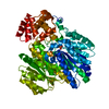


 PDBj
PDBj
 Assembly
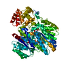
 TRICHOPLUSIA NI (cabbage looper) / References: UniProt: Q14397
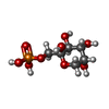
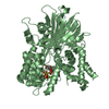
 Type: D-saccharide / Mass: 260.136 Da / Num. of mol.: 1
 Mass: 40.078 Da / Num. of mol.: 1 / Source method: obtained synthetically / Formula: Ca
 Mass: 18.015 Da / Num. of mol.: 700 / Source method: isolated from a natural source / Formula: H2O
 Sample preparation
 / Beamline: X06SA / Wavelength: 0.96
  Processing