Mass: 18.015 Da / Num. of mol.: 82 / Source method: isolated from a natural source / Formula: H2O
Has protein modification
Y
Nonpolymer details
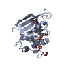
COBALT (II) ION (CO): CO REPLACES THE NATIVE FE HYDROSULFURIC ACID (H2S): H2S IS BOUND AS ...COBALT (II) ION (CO): CO REPLACES THE NATIVE FE HYDROSULFURIC ACID (H2S): H2S IS BOUND AS HYDROSULFIDE CYSTEINE SULFENIC ACID (CSO): NO FUNCTION ASSIGNED
Sequence details
C-TERMINAL HIS TAG
-
Experimental details
-
Experiment
Experiment
Method: X-RAY DIFFRACTION / Number of used crystals: 1
-
Sample preparation
Crystal
Density Matthews: 2.67 Å3/Da / Density % sol: 54 % / Description: NONE
Crystal grow
Temperature: 293 K / pH: 4.6 Details: 2 UL PROTEIN PLUS 2 UL (20% PEG 4000, 0.1 M NAOAC PH 4.6), 293 K. SOAKED IN 10% PEG4000, 20% PEG400, 0.1 M NAOAC PH 4.6, 293 K, FOR 1 DAY. INCUBATED IN H2S ATMOSPHERE EQUILIBRATED WITH 50 MM NA2S AT PH 4.6
Resolution: 1.8→46.38 Å / Cor.coef. Fo:Fc: 0.963 / Cor.coef. Fo:Fc free: 0.95 / SU B: 5.998 / SU ML: 0.082 / Cross valid method: THROUGHOUT / ESU R: 0.114 / ESU R Free: 0.111 / Stereochemistry target values: MAXIMUM LIKELIHOOD / Details: HYDROGENS HAVE BEEN ADDED IN THE RIDING POSITIONS.
Rfactor
Num. reflection
% reflection
Selection details
Rfree
0.22115
1014
5.1 %
RANDOM
Rwork
0.18874
-
-
-
obs
0.19035
18880
99.7 %
-
Solvent computation
Ion probe radii: 0.8 Å / Shrinkage radii: 0.8 Å / VDW probe radii: 1.2 Å / Solvent model: MASK
 Movie
Movie Controller


 Open data
 Basic information
 Components
 Keywords
 Function and homology information
 X-RAY DIFFRACTION /
  Authors
 Citation
 Structure visualization
 Downloads & links
 Other downloads


 PDBj
PDBj
 Assembly

 Mass: 58.933 Da / Num. of mol.: 1 / Source method: obtained synthetically / Formula: Co
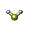
 Mass: 34.081 Da / Num. of mol.: 1 / Source method: obtained synthetically / Formula: H2S
 Mass: 18.015 Da / Num. of mol.: 82 / Source method: isolated from a natural source / Formula: H2O
 Sample preparation
 / Beamline: 14.1 / Wavelength: 0.918
  Processing